Chapman Christopher H, Zhu Tong, Nazem-Zadeh Mohamad, Tao Yebin, Buchtel Henry A, Tsien Christina I, Lawrence Theodore S, Cao Yue
Department of Radiation Oncology, University of Michigan, Ann Arbor, USA; Department of Radiation Oncology, University of California San Francisco, USA.
Department of Radiation Oncology, University of Michigan, Ann Arbor, USA.
Radiother Oncol. 2016 Aug;120(2):234-40. doi: 10.1016/j.radonc.2016.06.021. Epub 2016 Jul 11.
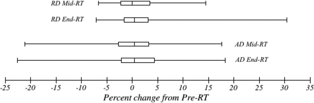
PURPOSE/OBJECTIVES: Radiation injury to parahippocampal cingulum white matter is associated with cognitive decline. Diffusion tensor imaging (DTI) detects micropathologic changes in white matter. Increased radial diffusion (RD) and decreased axial diffusion (AD) correspond to demyelination and axonal degeneration/gliosis respectively. We aimed to develop a predictive model for radiation-induced cognitive changes based upon DTI changes.
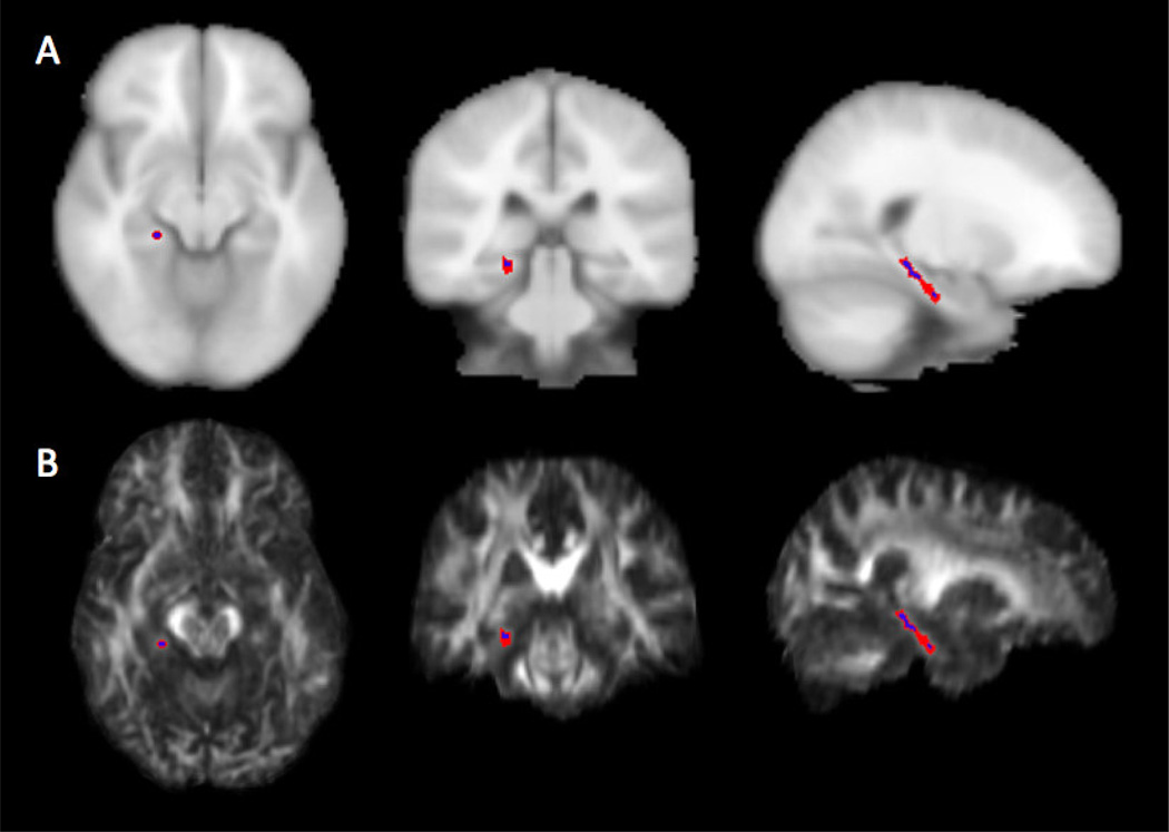
MATERIALS/METHODS: Twenty-seven adults with benign or low-grade tumors received partial brain radiation therapy (RT) to a median dose of 54Gy. Patients underwent DTI before RT, during RT, and at the end of RT. Cognitive testing was performed before RT, and 6 and 18months after RT. Parahippocampal cingulum white matter was contoured to obtain mean values of AD and RD.
By univariate analysis, decreasing AD and increasing RD during RT predicted declines in verbal memory and verbal fluency. By multivariate analysis, baseline neurocognitive score was the only clinical variable predicting verbal memory change; no clinical variables predicted verbal fluency change. In a multivariate model, increased RD at the end of RT significantly predicted decline in verbal fluency 18months after RT.
Imaging biomarkers of white matter injury contributed to predictive models of cognitive function change after RT.
目的/目标:海马旁扣带回白质的放射性损伤与认知功能下降有关。扩散张量成像(DTI)可检测白质中的微观病理变化。径向扩散(RD)增加和轴向扩散(AD)减少分别对应于脱髓鞘和轴突退变/胶质增生。我们旨在基于DTI变化开发一种预测放射性认知变化的模型。
材料/方法:27例患有良性或低度肿瘤的成年人接受了部分脑放射治疗(RT),中位剂量为54Gy。患者在放疗前、放疗期间和放疗结束时接受了DTI检查。在放疗前、放疗后6个月和18个月进行了认知测试。勾勒出海马旁扣带回白质轮廓以获得AD和RD的平均值。
单因素分析显示,放疗期间AD降低和RD升高预示着言语记忆和言语流畅性下降。多因素分析显示,基线神经认知评分是预测言语记忆变化的唯一临床变量;没有临床变量可预测言语流畅性变化。在多变量模型中,放疗结束时RD升高显著预测了放疗后18个月言语流畅性下降。
白质损伤的影像学生物标志物有助于建立放疗后认知功能变化的预测模型。