• 文献检索
  • 文档翻译
  • 深度研究
  • 学术资讯
  • Suppr Zotero 插件Zotero 插件
  • 邀请有礼
  • 套餐&价格
  • 历史记录
应用&插件
Suppr Zotero 插件Zotero 插件浏览器插件Mac 客户端Windows 客户端微信小程序
定价
高级版会员购买积分包购买API积分包
服务
文献检索文档翻译深度研究API 文档MCP 服务
关于我们
关于 Suppr公司介绍联系我们用户协议隐私条款
关注我们

Suppr 超能文献

核心技术专利:CN118964589B侵权必究
粤ICP备2023148730 号-1Suppr @ 2026

文献检索

告别复杂PubMed语法,用中文像聊天一样搜索,搜遍4000万医学文献。AI智能推荐,让科研检索更轻松。

立即免费搜索

文件翻译

保留排版,准确专业,支持PDF/Word/PPT等文件格式,支持 12+语言互译。

免费翻译文档

深度研究

AI帮你快速写综述,25分钟生成高质量综述,智能提取关键信息,辅助科研写作。

立即免费体验

7只犬肝脏腔静脉孔疝的影像学和计算机断层扫描特征:酷似肺结节

Radiographic and computed tomographic features of caval foramen hernias of the liver in 7 dogs: mimicking lung nodules.

作者信息

Kim Jaehwan, Kim Soyoung, Jo Jieun, Lee Seungjun, Eom Kidong

机构信息

Konkuk University, 120, Neungdong-ro, Gwangjin-gu, Seoul, Korea.

出版信息

J Vet Med Sci. 2016 Dec 1;78(11):1693-1697. doi: 10.1292/jvms.16-0161. Epub 2016 Jul 16.

DOI:10.1292/jvms.16-0161
PMID:27430293
原文链接:https://pmc.ncbi.nlm.nih.gov/articles/PMC5138423/
Abstract

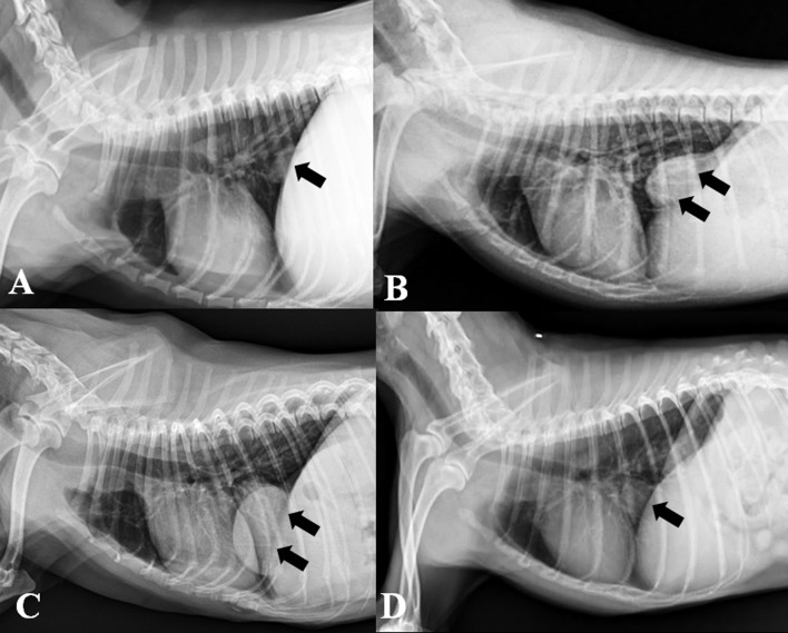
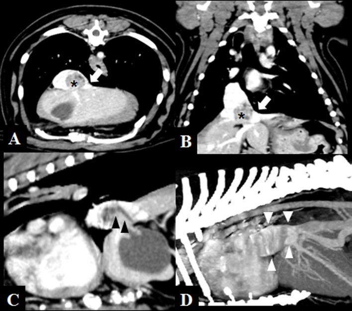
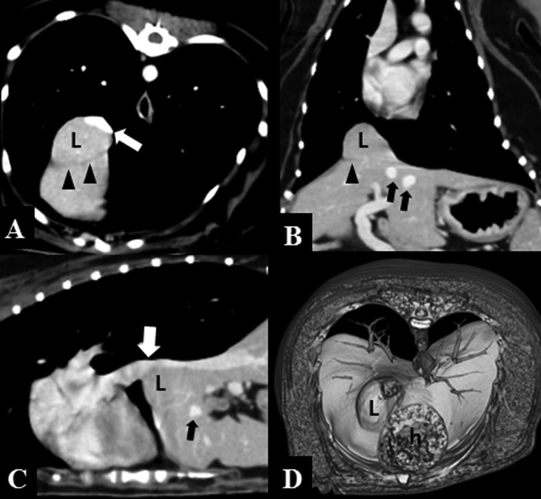
This study describes the imaging features and characteristics of caval foramen hernias in 7 dogs diagnosed by computed tomography (CT). On lateral radiographs, 6 of 7 dogs showed dome-shaped, broad-based, caudal mediastinal lesions. CT findings included caudal vena cava (CVC) compression (n=7), right lateral (n=6) or medial (n=1) liver lobe involvement, hepatic vein dilation (n=5) and biliary tract involvement (n=1) with partial (n=6) or entire (n=1) liver lobe hernias. A caval foramen hernia should be part of the differential diagnosis when the aforementioned imaging features are detected. CT is considered as a useful tool for diagnosis and evaluation in dogs with a caval foramen hernia.

摘要

本研究描述了7只经计算机断层扫描(CT)诊断为腔静脉孔疝的犬的影像学特征。在侧位X线片上,7只犬中有6只显示出圆顶状、宽基底的后纵隔病变。CT表现包括尾腔静脉(CVC)受压(n = 7)、右侧(n = 6)或内侧(n = 1)肝叶受累、肝静脉扩张(n = 5)以及胆道受累(n = 1),伴有部分(n = 6)或整个(n = 1)肝叶疝。当检测到上述影像学特征时,腔静脉孔疝应作为鉴别诊断的一部分。CT被认为是诊断和评估犬腔静脉孔疝的有用工具。

https://cdn.ncbi.nlm.nih.gov/pmc/blobs/ecd0/5138423/48695585305b/jvms-78-1693-g003.jpg
https://cdn.ncbi.nlm.nih.gov/pmc/blobs/ecd0/5138423/13462cfedf33/jvms-78-1693-g001.jpg
https://cdn.ncbi.nlm.nih.gov/pmc/blobs/ecd0/5138423/85ffc8e28bd7/jvms-78-1693-g002.jpg
https://cdn.ncbi.nlm.nih.gov/pmc/blobs/ecd0/5138423/48695585305b/jvms-78-1693-g003.jpg
https://cdn.ncbi.nlm.nih.gov/pmc/blobs/ecd0/5138423/13462cfedf33/jvms-78-1693-g001.jpg
https://cdn.ncbi.nlm.nih.gov/pmc/blobs/ecd0/5138423/85ffc8e28bd7/jvms-78-1693-g002.jpg
https://cdn.ncbi.nlm.nih.gov/pmc/blobs/ecd0/5138423/48695585305b/jvms-78-1693-g003.jpg

相似文献

1
Radiographic and computed tomographic features of caval foramen hernias of the liver in 7 dogs: mimicking lung nodules.7只犬肝脏腔静脉孔疝的影像学和计算机断层扫描特征:酷似肺结节
J Vet Med Sci. 2016 Dec 1;78(11):1693-1697. doi: 10.1292/jvms.16-0161. Epub 2016 Jul 16.
2
Caval foramen hernia in a dog: Preoperative diagnosis and surgical treatment.犬的腔静脉孔疝:术前诊断和手术治疗。
J Vet Med Sci. 2020 Nov 12;82(11):1602-1606. doi: 10.1292/jvms.19-0575. Epub 2020 Aug 14.
3
Radiographic diagnosis of diaphragmatic hernia: review of 60 cases in dogs and cats.膈疝的影像学诊断:犬猫60例病例回顾
J Vet Sci. 2004 Jun;5(2):157-62.
4
Computed tomographic features for differentiating benign from malignant liver lesions in dogs.犬肝脏良性与恶性病变鉴别的计算机断层扫描特征
J Vet Med Sci. 2019 Dec 18;81(12):1697-1704. doi: 10.1292/jvms.19-0278. Epub 2019 Oct 10.
5
Computed tomographic features of pneumothorax secondary to a bronchopleural fistula in two dogs.两只犬支气管胸膜瘘继发气胸的计算机断层扫描特征
J Am Anim Hosp Assoc. 2014 Jul-Aug;50(4):284-90. doi: 10.5326/JAAHA-MS-6010.
6
Radiographic, computed tomographic, and ultrasonographic findings with migrating intrathoracic grass awns in dogs and cats.犬猫体内迁移性胸内草芒的X线、计算机断层扫描及超声检查结果
Vet Radiol Ultrasound. 2008 May-Jun;49(3):249-55. doi: 10.1111/j.1740-8261.2008.00360.x.
7
Imaging diagnosis--positive contrast peritoneographic features of true diaphragmatic hernia.影像诊断——真性膈疝的阳性对比剂腹膜造影特征
Vet Radiol Ultrasound. 2009 Mar-Apr;50(2):185-7. doi: 10.1111/j.1740-8261.2009.01514.x.
8
What is your diagnosis? Peritoneopericardial diaphragmatic hernia.你的诊断是什么?腹膜心包膈疝。
J Am Vet Med Assoc. 2011 Apr 1;238(7):847-8. doi: 10.2460/javma.238.7.847.
9
Computed tomographic and magnetic resonance imaging features of canine segmental caudal vena cava aplasia.犬节段性尾腔静脉发育不全的计算机断层扫描和磁共振成像特征。
J Small Anim Pract. 2009 Jul;50(7):341-9. doi: 10.1111/j.1748-5827.2009.00748.x.
10
Computed tomographic morphology and clinical features of extrahepatic portosystemic shunts in 172 dogs in Japan.日本172只犬肝外门体分流的计算机断层扫描形态及临床特征
Vet J. 2014 Mar;199(3):376-81. doi: 10.1016/j.tvjl.2013.11.013. Epub 2013 Nov 23.

引用本文的文献

1
Successful early diagnosis and surgical treatment of congenital caval foramen hernia in an 8-month-old mixed breed cat.8个月大的混种猫先天性腔静脉孔疝的早期成功诊断与手术治疗
Braz J Vet Med. 2023 Jan 27;45:e005622. doi: 10.29374/2527-2179.bjvm005622. eCollection 2023.
2
Caval foramen hernia in a cat.猫的腔静脉孔疝
JFMS Open Rep. 2020 Oct 21;6(2):2055116920964021. doi: 10.1177/2055116920964021. eCollection 2020 Jul-Dec.
3
Caval foramen hernia in a dog: Preoperative diagnosis and surgical treatment.犬的腔静脉孔疝:术前诊断和手术治疗。

本文引用的文献

1
Hernia of the diaphragmatic caval foramen causing right atrial "mass", caval obstruction and pulmonary embolism.膈肌腔静脉孔疝导致右心房“肿块”、腔静脉阻塞和肺栓塞。
Int J Cardiol. 2016 Mar 15;207:215-6. doi: 10.1016/j.ijcard.2016.01.166. Epub 2016 Jan 11.
2
Imaging evaluation of the inferior vena cava.下腔静脉的影像学评估。
Radiographics. 2015 Mar-Apr;35(2):578-92. doi: 10.1148/rg.352140136.
3
Retrosternal (Morgagni) diaphragmatic hernia.胸骨后(莫尔加尼)膈疝。
J Vet Med Sci. 2020 Nov 12;82(11):1602-1606. doi: 10.1292/jvms.19-0575. Epub 2020 Aug 14.
Can Vet J. 2011 Aug;52(8):878-83.
4
Budd-Chiari-like syndrome in a dog due to liver lobe entrapment within the falciform ligament.一只犬因镰状韧带内肝叶嵌顿导致布-加样综合征。
J Am Anim Hosp Assoc. 2009 Sep-Oct;45(5):253-6. doi: 10.5326/0450253.
5
Imaging diagnosis--positive contrast peritoneographic features of true diaphragmatic hernia.影像诊断——真性膈疝的阳性对比剂腹膜造影特征
Vet Radiol Ultrasound. 2009 Mar-Apr;50(2):185-7. doi: 10.1111/j.1740-8261.2009.01514.x.
6
Imaging the inferior vena cava: a road less traveled.下腔静脉成像:一条少有人走的路。
Radiographics. 2008 May-Jun;28(3):669-89. doi: 10.1148/rg.283075101.
7
Budd-Chiari-like syndrome caused by a congenital hiatal hernia in a shar-pei dog.一只沙皮犬因先天性食管裂孔疝引起的布-加氏综合征样综合征。
Vet Rec. 2006 Sep 2;159(10):322-3. doi: 10.1136/vr.159.10.322.
8
Caval foramen hernia masquerading as a thoracic mass.伪装成胸部肿块的腔静脉孔疝
Can J Surg. 2006 Feb;49(1):64-5.
9
Eventration of the diaphragm.膈肌膨出
Asian J Surg. 2006 Jan;29(1):8-10. doi: 10.1016/S1015-9584(09)60285-2.
10
Traumatic diaphragmatic hernia in a clinically normal dog.一只临床健康犬的创伤性膈疝
Can Vet J. 2001 Jul;42(7):564-6.