Kvitka Dmitrij, Juodžentė Dalia, Rudenkovaitė Greta, Burbaitė Evelina, Laukutė Monika
Veterinarian, Dr. L. Kriaučeliūnas Small Animal Veterinary Clinic, Faculty of Veterinary, Veterinary Academy, Lithuanian University of Health Sciences, Kaunas, Lithuania.
Veterinarian, Neurology and Neurosurgery Division, San Marco Veterinary Clinic, Veggiano, Italy.
Braz J Vet Med. 2023 Jan 27;45:e005622. doi: 10.29374/2527-2179.bjvm005622. eCollection 2023.
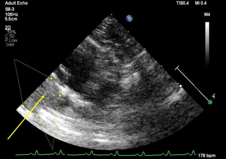
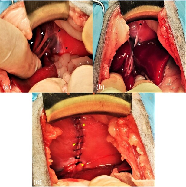
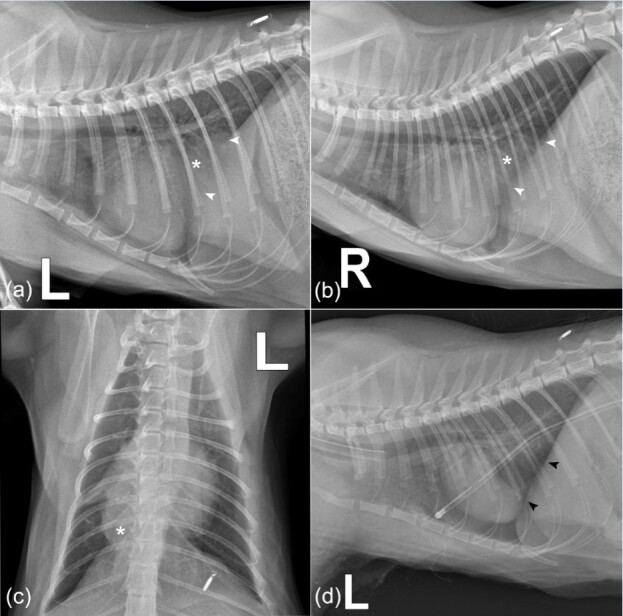
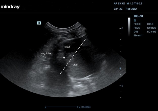
An 8-month-old neutered female domestic mixed breed cat was presented to Dr. L. Kriaučeliūnas Small Animal Clinic due to coughing that persisted for 2 weeks. Lateral and dorsoventral chest radiographs revealed an unusual dome-shaped soft tissue opacity mass that had contact with the cranial part of the diaphragm. Together with heart and abdominal ultrasound findings, we decided that one of the differential diagnoses was a diaphragmatic hernia. During the diagnostic celiotomy, a vertical 4 cm in length diaphragmatic deficit was visualized. Left medial and lateral liver lobes were herniated, yet healthy-looking. Adhesions between the liver lobes and the pericardium sac were visualized and dissected. The pericardium was sutured with simple interrupted suture pattern. A herniorrhaphy was performed suturing the diaphragm with the continuous suture pattern. Successful surgical treatment resulted in fully resolved clinical symptoms.
一只8个月大已绝育的雌性家猫混种猫因持续咳嗽2周被送至L. Kriaučeliūnas小动物诊所。胸部侧位和背腹位X光片显示一个异常的圆顶形软组织密度肿块,与膈肌的头端部分接触。结合心脏和腹部超声检查结果,我们确定鉴别诊断之一为膈疝。在诊断性剖腹术中,可见一个长4厘米的垂直膈肌缺损。左内侧和外侧肝叶发生疝出,但外观健康。可见并分离了肝叶与心包囊之间的粘连。心包用单纯间断缝合法缝合。采用连续缝合法对膈肌进行疝修补术。成功的手术治疗使临床症状完全缓解。