Domanski Dominik, Perzanowska Anna, Kistowski Michal, Wojtas Grzegorz, Michalak Agata, Krasowski Grzegorz, Dadlez Michal
Institute of Biochemistry and Biophysics, Polish Academy of Sciences, Pawinskiego 5A, 02-106 Warsaw, Poland.
Institute of Biochemistry and Biophysics, Polish Academy of Sciences, Pawinskiego 5A, 02-106 Warsaw, Poland.
Neoplasia. 2016 Jul;18(7):399-412. doi: 10.1016/j.neo.2016.06.002. Epub 2016 Jun 25.
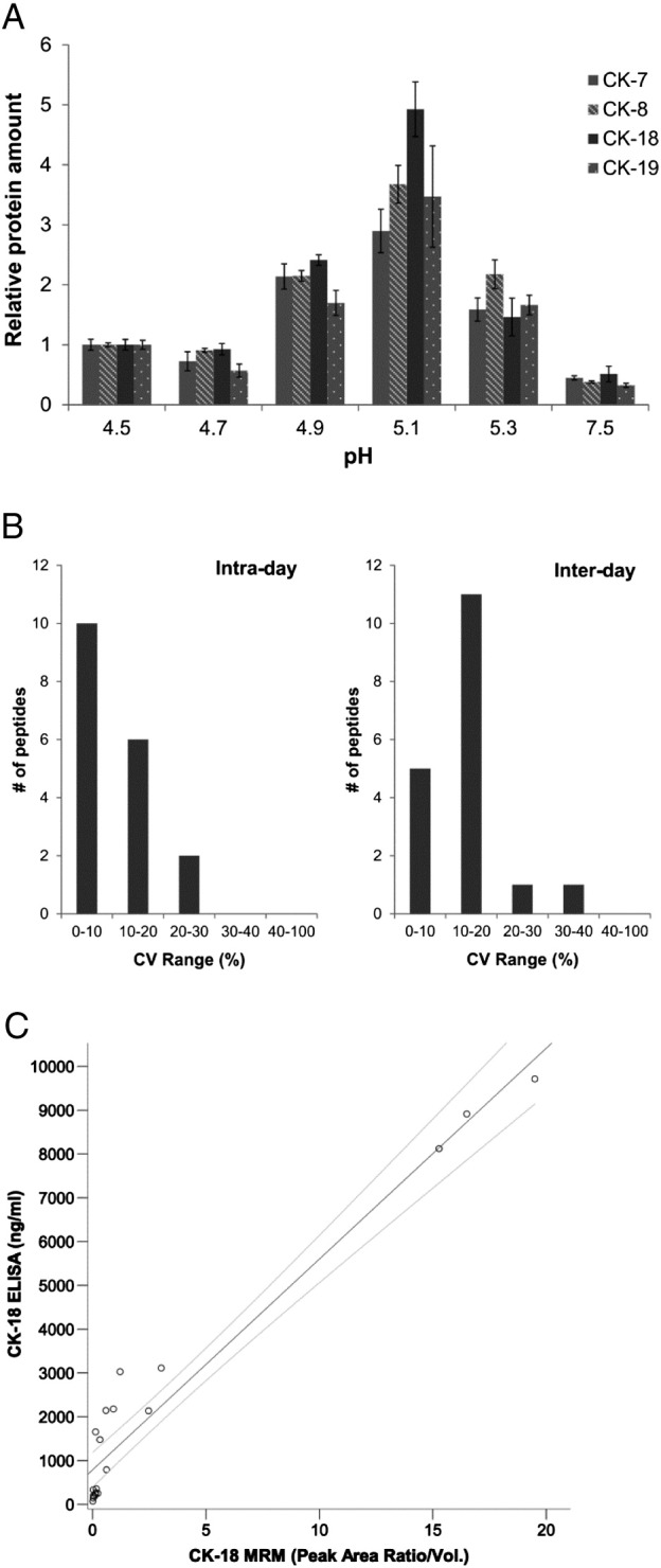
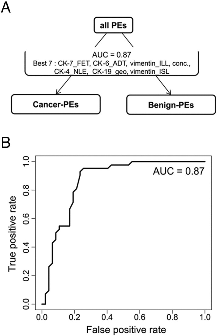
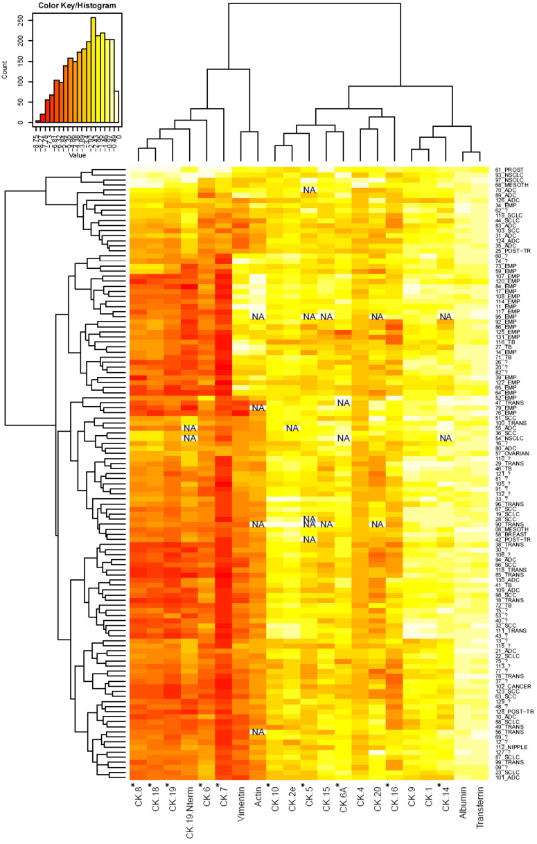
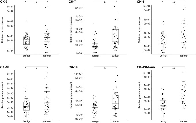
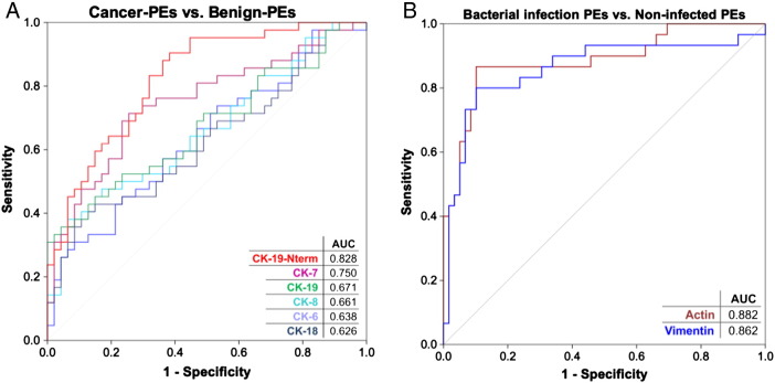
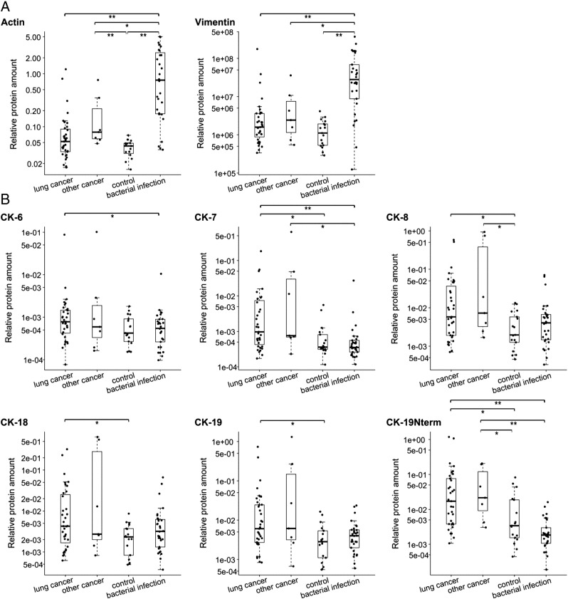
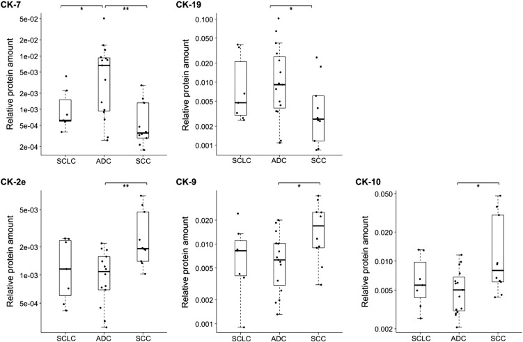
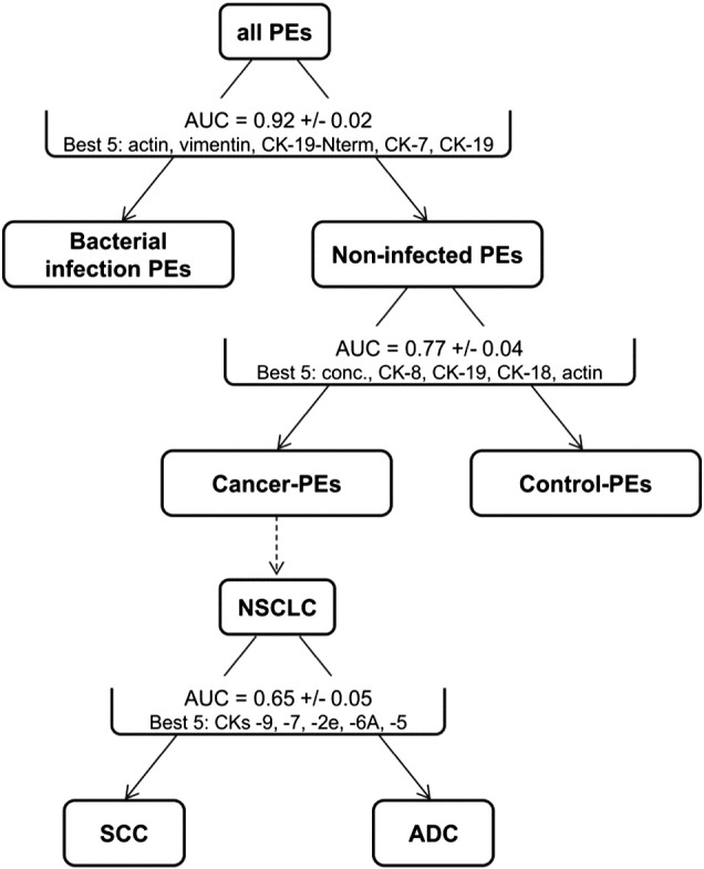
Pleural effusion (PE), excess fluid in the pleural space, is often observed in lung cancer patients and also forms due to many benign ailments. Classifying it quickly is critical, but this remains an analytical challenge often lengthening the diagnosis process or exposing patients to unnecessary risky invasive procedures. We tested the analysis of PE using a multiplexed cytokeratin (CK) panel with targeted mass spectrometry-based quantitation for its rapid classification. CK markers are often assessed in pathological examinations for cancer diagnosis and guiding treatment course. We developed methods to simultaneously quantify 33 CKs in PE using peptide standards for increased analytical specificity and a simple CK enrichment method to detect their low amounts. Analyzing 121 PEs associated with a variety of lung cancers and noncancerous causes, we show that abundance levels of 10 CKs can be related to PE etiology. CK-6, CK-7, CK-8, CK-18, and CK-19 were found at significantly higher levels in cancer-related PEs. Additionally, elevated levels of vimentin and actin differentiated PEs associated with bacterial infections. A classifier algorithm effectively grouped PEs into cancer-related or benign PEs with 81% sensitivity and 79% specificity. A set of undiagnosed PEs showed that our method has potential to shorten PE diagnosis time. For the first time, we show that a cancer-relevant panel of simple-epithelial CK markers currently used in clinical assessment can also be quantitated in PEs. Additionally, while requiring less invasive sampling, our methodology demonstrated a significant ability to identify cancer-related PEs in clinical samples and thus could improve patient care in the future.
胸腔积液(PE)是指胸腔内出现过多液体,在肺癌患者中较为常见,也可由多种良性疾病引起。快速对其进行分类至关重要,但这仍然是一项分析挑战,常常会延长诊断过程,或者使患者面临不必要的有风险的侵入性检查。我们使用多重细胞角蛋白(CK)检测板并结合基于靶向质谱的定量分析来测试胸腔积液的分析,以实现快速分类。CK标志物常用于病理检查以诊断癌症并指导治疗过程。我们开发了方法,使用肽标准品同时定量胸腔积液中的33种CK,以提高分析特异性,并采用一种简单的CK富集方法来检测其低含量。通过分析121例与各种肺癌和非癌病因相关的胸腔积液,我们发现10种CK的丰度水平与胸腔积液的病因有关。在与癌症相关的胸腔积液中,CK-6、CK-7、CK-8、CK-18和CK-19的水平显著更高。此外,波形蛋白和肌动蛋白水平升高可区分与细菌感染相关的胸腔积液。一种分类算法能有效地将胸腔积液分为癌症相关或良性胸腔积液,灵敏度为81%,特异性为79%。一组未确诊的胸腔积液表明我们的方法有缩短胸腔积液诊断时间的潜力。我们首次表明,目前临床评估中使用的一组与癌症相关的简单上皮CK标志物也可在胸腔积液中进行定量。此外,虽然需要的侵入性采样较少,但我们的方法在识别临床样本中与癌症相关的胸腔积液方面显示出显著能力,因此未来可能会改善患者护理。