• 文献检索
  • 文档翻译
  • 深度研究
  • 学术资讯
  • Suppr Zotero 插件Zotero 插件
  • 邀请有礼
  • 套餐&价格
  • 历史记录
应用&插件
Suppr Zotero 插件Zotero 插件浏览器插件Mac 客户端Windows 客户端微信小程序
定价
高级版会员购买积分包购买API积分包
服务
文献检索文档翻译深度研究API 文档MCP 服务
关于我们
关于 Suppr公司介绍联系我们用户协议隐私条款
关注我们

Suppr 超能文献

核心技术专利:CN118964589B侵权必究
粤ICP备2023148730 号-1Suppr @ 2026

文献检索

告别复杂PubMed语法,用中文像聊天一样搜索,搜遍4000万医学文献。AI智能推荐,让科研检索更轻松。

立即免费搜索

文件翻译

保留排版,准确专业,支持PDF/Word/PPT等文件格式,支持 12+语言互译。

免费翻译文档

深度研究

AI帮你快速写综述,25分钟生成高质量综述,智能提取关键信息,辅助科研写作。

立即免费体验

用于评估肾纤维化的新型磁共振成像指标:扩散加权成像与T1映射成像的比较及组织学验证

New Magnetic Resonance Imaging Index for Renal Fibrosis Assessment: A Comparison between Diffusion-Weighted Imaging and T1 Mapping with Histological Validation.

作者信息

Friedli I, Crowe L A, Berchtold L, Moll S, Hadaya K, de Perrot T, Vesin C, Martin P-Y, de Seigneux S, Vallée J-P

机构信息

Division of Radiology, Department of Radiology and Medical Informatics Geneva University Hospitals and Faculty of Medicine of the University of Geneva, Switzerland.

Service of Nephrology, Department of Internal Medicine Specialties, Geneva University Hospitals, University of Geneva, Faculty of Medicine, Geneva, Switzerland.

出版信息

Sci Rep. 2016 Jul 21;6:30088. doi: 10.1038/srep30088.

DOI:10.1038/srep30088
PMID:27439482
原文链接:https://pmc.ncbi.nlm.nih.gov/articles/PMC4954968/
Abstract

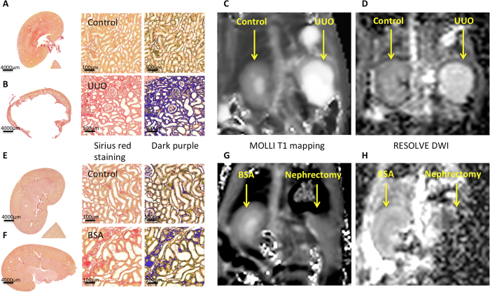
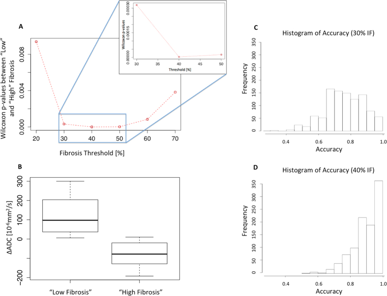
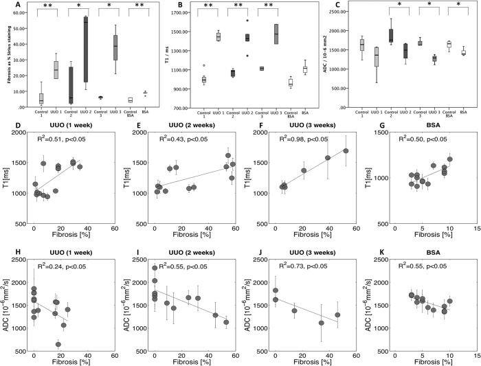
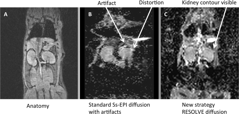
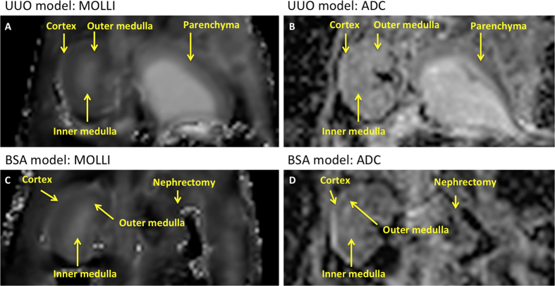
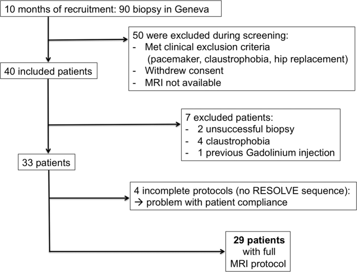
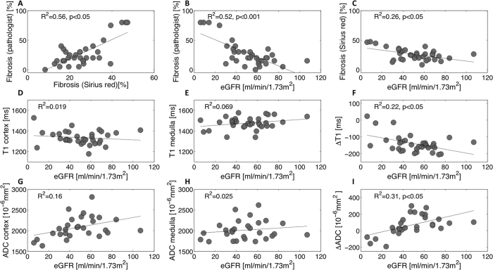
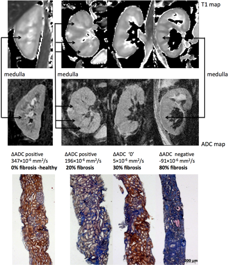
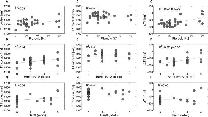
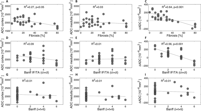
A need exists to noninvasively assess renal interstitial fibrosis, a common process to all kidney diseases and predictive of renal prognosis. In this translational study, Magnetic Resonance Imaging (MRI) T1 mapping and a new segmented Diffusion-Weighted Imaging (DWI) technique, for Apparent Diffusion Coefficient (ADC), were first compared to renal fibrosis in two well-controlled animal models to assess detection limits. Validation against biopsy was then performed in 33 kidney allograft recipients (KARs). Predictive MRI indices, ΔT1 and ΔADC (defined as the cortico-medullary differences), were compared to histology. In rats, both T1 and ADC correlated well with fibrosis and inflammation showing a difference between normal and diseased kidneys. In KARs, MRI indices were not sensitive to interstitial inflammation. By contrast, ΔADC outperformed ΔT1 with a stronger negative correlation to fibrosis (R(2) = 0.64 against R(2) = 0.29 p < 0.001). ΔADC tends to negative values in KARs harboring cortical fibrosis of more than 40%. Using a discriminant analysis method, the ΔADC, as a marker to detect such level of fibrosis or higher, led to a specificity and sensitivity of 100% and 71%, respectively. This new index has potential for noninvasive assessment of fibrosis in the clinical setting.

摘要

需要对肾间质纤维化进行无创评估,肾间质纤维化是所有肾脏疾病的常见过程,且可预测肾脏预后。在这项转化研究中,首先在两种严格控制的动物模型中,将磁共振成像(MRI)T1映射和一种用于表观扩散系数(ADC)的新型分段扩散加权成像(DWI)技术与肾纤维化进行比较,以评估检测限。然后在33名肾移植受者(KAR)中进行了与活检的验证。将预测性MRI指标ΔT1和ΔADC(定义为皮质 - 髓质差异)与组织学进行比较。在大鼠中,T1和ADC均与纤维化和炎症密切相关,显示出正常肾脏和患病肾脏之间的差异。在KAR中,MRI指标对间质炎症不敏感。相比之下,ΔADC优于ΔT1,与纤维化的负相关性更强(R² = 0.64,而R² = 0.29,p < 0.001)。在皮质纤维化超过40%的KAR中,ΔADC倾向于负值。使用判别分析方法,ΔADC作为检测这种纤维化水平或更高水平的标志物,特异性和敏感性分别为100%和71%。这一新指标在临床环境中具有无创评估纤维化的潜力。

https://cdn.ncbi.nlm.nih.gov/pmc/blobs/02de/4954968/54c38a57045b/srep30088-f10.jpg
https://cdn.ncbi.nlm.nih.gov/pmc/blobs/02de/4954968/a2d51253d461/srep30088-f1.jpg
https://cdn.ncbi.nlm.nih.gov/pmc/blobs/02de/4954968/6d9e7581bb6c/srep30088-f2.jpg
https://cdn.ncbi.nlm.nih.gov/pmc/blobs/02de/4954968/90c6ca7ef629/srep30088-f3.jpg
https://cdn.ncbi.nlm.nih.gov/pmc/blobs/02de/4954968/19c2b8a9424c/srep30088-f4.jpg
https://cdn.ncbi.nlm.nih.gov/pmc/blobs/02de/4954968/b58a7a686b7a/srep30088-f5.jpg
https://cdn.ncbi.nlm.nih.gov/pmc/blobs/02de/4954968/2ab675e23df2/srep30088-f6.jpg
https://cdn.ncbi.nlm.nih.gov/pmc/blobs/02de/4954968/dd964b91b8d2/srep30088-f7.jpg
https://cdn.ncbi.nlm.nih.gov/pmc/blobs/02de/4954968/902c0fcb5de7/srep30088-f8.jpg
https://cdn.ncbi.nlm.nih.gov/pmc/blobs/02de/4954968/c29d8d8c8dbc/srep30088-f9.jpg
https://cdn.ncbi.nlm.nih.gov/pmc/blobs/02de/4954968/54c38a57045b/srep30088-f10.jpg

相似文献

1
New Magnetic Resonance Imaging Index for Renal Fibrosis Assessment: A Comparison between Diffusion-Weighted Imaging and T1 Mapping with Histological Validation.用于评估肾纤维化的新型磁共振成像指标:扩散加权成像与T1映射成像的比较及组织学验证
Sci Rep. 2016 Jul 21;6:30088. doi: 10.1038/srep30088.
2
Validation of the corticomedullary difference in magnetic resonance imaging-derived apparent diffusion coefficient for kidney fibrosis detection: a cross-sectional study.基于磁共振成像表观扩散系数的皮质-髓质差异对肾脏纤维化检测的验证:一项横断面研究。
Nephrol Dial Transplant. 2020 Jun 1;35(6):937-945. doi: 10.1093/ndt/gfy389.
3
Comparison of readout-segmented and conventional single-shot for echo-planar diffusion-weighted imaging in the assessment of kidney interstitial fibrosis.在评估肾间质纤维化中,读出分段和常规单次激发 echo-planar 扩散加权成像的比较。
J Magn Reson Imaging. 2017 Dec;46(6):1631-1640. doi: 10.1002/jmri.25687. Epub 2017 Mar 10.
4
Validation of automatically measured T1 map cortico-medullary difference (ΔT1) for eGFR and fibrosis assessment in allograft kidneys.验证自动测量的皮质-髓质 T1 映射差值 (ΔT1) 用于评估同种异体移植肾脏的 eGFR 和纤维化。
PLoS One. 2023 Feb 15;18(2):e0277277. doi: 10.1371/journal.pone.0277277. eCollection 2023.
5
Multiparametric magnetic resonance imaging shows promising results to assess renal transplant dysfunction with fibrosis.多参数磁共振成像在评估伴有纤维化的肾移植功能障碍方面显示出有希望的结果。
Kidney Int. 2020 Feb;97(2):414-420. doi: 10.1016/j.kint.2019.09.030. Epub 2019 Oct 30.
6
Diffusion-weighted MRI does not reflect kidney fibrosis in a rat model of fibrosis.弥散加权 MRI 不能反映纤维化大鼠模型中的肾脏纤维化。
J Magn Reson Imaging. 2015 Oct;42(4):990-8. doi: 10.1002/jmri.24853. Epub 2015 Jan 28.
7
T2 relaxation time and apparent diffusion coefficient for noninvasive assessment of renal pathology after acute kidney injury in mice: comparison with histopathology.T2 弛豫时间和表观扩散系数在急性肾损伤后小鼠肾脏病变的无创评估中的应用:与组织病理学的比较。
Invest Radiol. 2013 Dec;48(12):834-42. doi: 10.1097/RLI.0b013e31829d0414.
8
Amide Proton Transfer-Weighted Magnetic Resonance Imaging for Application in Renal Fibrosis: A Radiological-Pathological-Based Analysis.酰胺质子转移加权磁共振成像在肾纤维化中的应用:基于放射病理的分析。
Am J Nephrol. 2024;55(3):334-344. doi: 10.1159/000536232. Epub 2024 Jan 16.
9
Diffusion-magnetic resonance imaging predicts decline of kidney function in chronic kidney disease and in patients with a kidney allograft.扩散加权磁共振成像可预测慢性肾脏病及肾移植患者的肾功能衰退。
Kidney Int. 2022 Apr;101(4):804-813. doi: 10.1016/j.kint.2021.12.014. Epub 2022 Jan 11.
10
Value of diffusion-weighted magnetic resonance imaging for prediction and early assessment of response to neoadjuvant radiochemotherapy in rectal cancer: preliminary results.弥散加权磁共振成像对直肠癌新辅助放化疗反应的预测及早期评估价值:初步结果。
Int J Radiat Oncol Biol Phys. 2012 Feb 1;82(2):863-70. doi: 10.1016/j.ijrobp.2010.12.063. Epub 2011 Mar 11.

引用本文的文献

1
The Possibilities of Multiparametric Magnetic Resonance Imaging to Reflect Functional and Structural Graft Changes 1 Year After Kidney Transplantation.多参数磁共振成像反映肾移植术后1年移植物功能和结构变化的可能性
Medicina (Kaunas). 2025 Jul 13;61(7):1268. doi: 10.3390/medicina61071268.
2
Prediction of early renal allograft dysfunction using multiparametric magnetic resonance imaging.使用多参数磁共振成像预测早期肾移植功能障碍
Quant Imaging Med Surg. 2025 May 1;15(5):4333-4342. doi: 10.21037/qims-24-2182. Epub 2025 Apr 22.
3
T Relaxation Time for the Prediction of Renal Transplant Dysfunction.

本文引用的文献

1
Diffusion in prostate cancer detection on a 3T scanner: How many b-values are needed?3T磁共振成像仪在前列腺癌检测中的扩散加权成像:需要多少个b值?
J Magn Reson Imaging. 2016 Sep;44(3):601-9. doi: 10.1002/jmri.25206. Epub 2016 Feb 23.
2
Kidney Transplantation: Multiparametric Functional Magnetic Resonance Imaging for Assessment of Renal Allograft Pathophysiology in Mice.肾移植:多参数功能磁共振成像评估小鼠肾移植病理生理学。
Invest Radiol. 2016 Jan;51(1):58-65. doi: 10.1097/RLI.0000000000000205.
3
What is the best way to measure renal fibrosis?: A pathologist's perspective.
用于预测肾移植功能障碍的T弛豫时间。
Transpl Int. 2025 Apr 10;38:14301. doi: 10.3389/ti.2025.14301. eCollection 2025.
4
T1 mapping magnetic resonance imaging predicts decline of kidney function.T1 映射磁共振成像可预测肾功能衰退。
Clin Kidney J. 2025 Feb 7;18(3):sfaf032. doi: 10.1093/ckj/sfaf032. eCollection 2025 Mar.
5
Utility of the combination of IVIM-DWI MRI and baseline eGFR for identifying a high risk of chronic kidney disease progression.IVIM-DWI磁共振成像与基线估算肾小球滤过率相结合在识别慢性肾病进展高风险方面的效用。
Front Med (Lausanne). 2025 Feb 19;12:1532210. doi: 10.3389/fmed.2025.1532210. eCollection 2025.
6
A first-in-human study of quantitative ultrasound to assess transplant kidney fibrosis.一项评估移植肾纤维化的定量超声人体首次研究。
Nat Med. 2025 Mar;31(3):970-978. doi: 10.1038/s41591-024-03417-5. Epub 2025 Mar 3.
7
Multiparametric MRI: can we assess renal function differently?多参数磁共振成像:我们能否以不同方式评估肾功能?
Clin Kidney J. 2024 Nov 19;18(1):sfae365. doi: 10.1093/ckj/sfae365. eCollection 2025 Jan.
8
Potential MRI Biomarkers for Predicting Kidney Function and Histological Damage in Transplanted Deceased Donor Kidney Recipients.预测已故供体肾移植受者肾功能和组织学损伤的潜在MRI生物标志物
J Clin Med. 2025 Feb 18;14(4):1349. doi: 10.3390/jcm14041349.
9
Assessment of renal insufficiency in patients with renal artery stenosis by multiparametric magnetic resonance imaging.通过多参数磁共振成像评估肾动脉狭窄患者的肾功能不全
Ren Fail. 2025 Dec;47(1):2444403. doi: 10.1080/0886022X.2024.2444403. Epub 2025 Jan 6.
10
Delta corticomedullary apparent diffusion coefficient on MRI as a biomarker for prognosis in IgA nephropathy.MRI上的皮质髓质Delta表观扩散系数作为IgA肾病预后的生物标志物
Ren Fail. 2025 Dec;47(1):2441394. doi: 10.1080/0886022X.2024.2441394. Epub 2024 Dec 17.
测量肾纤维化的最佳方法是什么?:病理学家的观点。
Kidney Int Suppl (2011). 2014 Nov;4(1):9-15. doi: 10.1038/kisup.2014.3.
4
Banff 2013 update: Pearls and pitfalls in transplant renal pathology.《班夫2013年更新版:移植肾病理学的要点与陷阱》
Nephrology (Carlton). 2015 Jul;20 Suppl 2:2-8. doi: 10.1111/nep.12474.
5
Improvement of renal diffusion-weighted magnetic resonance imaging with readout-segmented echo-planar imaging at 3T.3T下采用读出分段回波平面成像技术改善肾脏扩散加权磁共振成像
Magn Reson Imaging. 2015 Jul;33(6):701-8. doi: 10.1016/j.mri.2015.02.023. Epub 2015 Feb 27.
6
Diffusion-weighted MRI does not reflect kidney fibrosis in a rat model of fibrosis.弥散加权 MRI 不能反映纤维化大鼠模型中的肾脏纤维化。
J Magn Reson Imaging. 2015 Oct;42(4):990-8. doi: 10.1002/jmri.24853. Epub 2015 Jan 28.
7
T1 mapping: characterisation of myocardial interstitial space.T1 映射:心肌间质空间的特征描述
Insights Imaging. 2015 Apr;6(2):189-202. doi: 10.1007/s13244-014-0366-9. Epub 2014 Nov 26.
8
Recent advances in renal interstitial fibrosis and tubular atrophy after kidney transplantation.肾移植后肾间质纤维化和肾小管萎缩的最新进展
Fibrogenesis Tissue Repair. 2014 Oct 2;7:15. doi: 10.1186/1755-1536-7-15. eCollection 2014.
9
Assessment of renal fibrosis in chronic kidney disease using diffusion-weighted MRI.使用扩散加权磁共振成像评估慢性肾脏病中的肾纤维化
Clin Radiol. 2014 Nov;69(11):1117-22. doi: 10.1016/j.crad.2014.06.011. Epub 2014 Jul 23.
10
T1-mapping for assessment of ischemia-induced acute kidney injury and prediction of chronic kidney disease in mice.利用T1映射评估小鼠缺血性急性肾损伤及预测慢性肾病
Eur Radiol. 2014 Sep;24(9):2252-60. doi: 10.1007/s00330-014-3250-6. Epub 2014 Jul 5.