Hong Jung-Hwa, Park Mikyoung
Center for Functional Connectomics, Korea Institute of Science and TechnologySeoul, South Korea; Department of Life Sciences, Korea UniversitySeoul, South Korea.
Center for Functional Connectomics, Korea Institute of Science and TechnologySeoul, South Korea; Department of Neuroscience, Korea University of Science and TechnologyDaejeon, South Korea.
Front Synaptic Neurosci. 2016 Jun 29;8:18. doi: 10.3389/fnsyn.2016.00018. eCollection 2016.
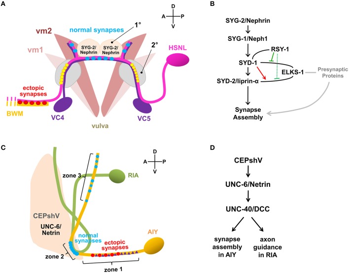
Formation of functional synapses is a fundamental process for establishing neural circuits and ultimately for expressing complex behavior. Extensive research has interrogated how such functional synapses are formed and how synapse formation contributes to the generation of neural circuitry and behavior. The nervous system of Caenorhabditis elegans, due to its relatively simple structure, the transparent body, and tractable genetic system, has been adapted as an excellent model to investigate synapses and the functional connectome. Advances in imaging technology together with the improvement of genetically encoded molecular tools enabled us to visualize synapses and neural circuits of the animal model, which provide insights into our understanding of molecules and their signaling pathways that mediate synapse formation and neuronal network modulation. Here, we review synaptogenesis in active zones and the mapping of local connectome in C. elegans nervous system whose understandings have been extended by the advances in imaging technology along with the genetic molecular tools.
功能性突触的形成是建立神经回路以及最终表现复杂行为的一个基本过程。广泛的研究探讨了这种功能性突触是如何形成的,以及突触形成如何促进神经回路和行为的产生。秀丽隐杆线虫的神经系统因其相对简单的结构、透明的身体和易于处理的遗传系统,已成为研究突触和功能连接组的优秀模型。成像技术的进步以及基因编码分子工具的改进使我们能够可视化动物模型的突触和神经回路,这为我们理解介导突触形成和神经网络调节的分子及其信号通路提供了见解。在这里,我们回顾了活性区的突触发生以及秀丽隐杆线虫神经系统中局部连接组的映射,成像技术和基因分子工具的进步扩展了我们对这些方面的理解。