Takasaki Chihiro, Kobayashi Masashi, Ishibashi Hironori, Akashi Takumi, Okubo Kenichi
Department of Thoracic Surgery, Tokyo Medical and Dental University, Tokyo 113-0034, Japan.
Department of Pathology, Tokyo Medical and Dental University, Tokyo 113-0034, Japan.
Mol Clin Oncol. 2016 Aug;5(2):295-300. doi: 10.3892/mco.2016.937. Epub 2016 Jun 17.
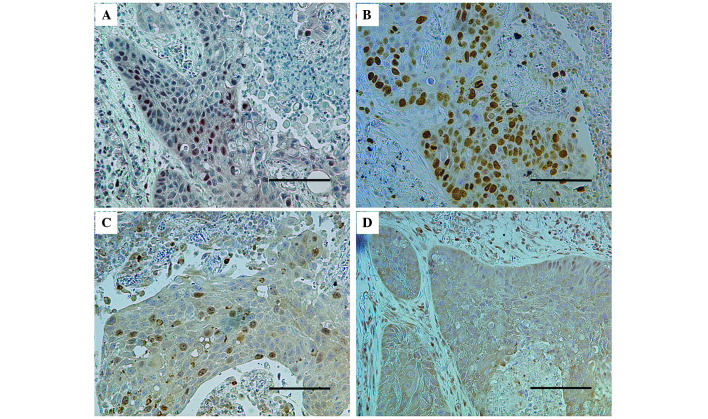
Hypoxia-inducible factor (HIF)-1 is a transcription factor that allows cells to adapt to hypoxic situations. HIF-1 is known to control tissue proliferation, antiapoptosis, angiogenesis and glucose metabolism. Furthermore, HIF-1 is involved in the growth of numerous cancer types. The present study aimed to examine the expression of HIF-1α immunohistochemically in resected lung cancers. The present study included 216 consecutive patients with lung cancer who underwent resection between April 2013 and January 2015. The patients' clinicopathological data were summarized, including imaging findings, tumor pathological characteristics, and the patient's age, sex and smoking status. The intratumoral expression of HIF-1α, survivin, c-Myc and the Ki-67 proliferation index were evaluated immunohistochemically. The patients were divided into two groups, according to the expression of HIF-1α (low vs. high) and the clinicopathological characteristics of these groups were compared. It was revealed that HIF-1α expression was significantly associated with ground glass opacity ratio, maximum standardized uptake value index, histological type (squamous cell carcinoma), differentiation and lymphatic invasion. Regarding the immunohistochemical findings, HIF-1α expression was significantly correlated with the expression levels of c-Myc (P<0.01) and survivin (P<0.01). Furthermore, the Ki-67 proliferation index was significantly higher in high-HIF-1α tumors compared with in low-HIF-1α tumors (P=0.01). The multivariate analysis identified squamous cell carcinoma, high SUVmax and lymphatic invasion as significant and independent factors for high HIF-1α expression. In conclusion, HIF-1 was highly expressed in certain subgroups of lung cancer with specific histopathology and images. HIF-1α expression was associated with tumor proliferation and antiapoptosis in lung cancer.
缺氧诱导因子(HIF)-1是一种转录因子,可使细胞适应缺氧环境。已知HIF-1能控制组织增殖、抗凋亡、血管生成和葡萄糖代谢。此外,HIF-1还参与多种癌症类型的生长。本研究旨在通过免疫组织化学方法检测HIF-1α在切除的肺癌中的表达情况。本研究纳入了2013年4月至2015年1月期间连续接受手术切除的216例肺癌患者。总结了患者的临床病理资料,包括影像学检查结果、肿瘤病理特征以及患者的年龄、性别和吸烟状况。通过免疫组织化学方法评估肿瘤内HIF-1α、生存素、c-Myc的表达以及Ki-67增殖指数。根据HIF-1α的表达情况(低表达与高表达)将患者分为两组,并比较两组的临床病理特征。结果显示,HIF-1α表达与磨玻璃影比例、最大标准化摄取值指数、组织学类型(鳞状细胞癌)、分化程度和淋巴管侵犯显著相关。关于免疫组织化学结果,HIF-1α表达与c-Myc(P<0.01)和生存素(P<0.01)的表达水平显著相关。此外,高HIF-1α表达的肿瘤中Ki-67增殖指数显著高于低HIF-1α表达的肿瘤(P=0.01)。多因素分析确定鳞状细胞癌、高SUVmax和淋巴管侵犯是HIF-1α高表达的显著独立因素。总之,HIF-1在具有特定组织病理学和影像学特征的肺癌亚组中高表达。HIF-1α表达与肺癌的肿瘤增殖和抗凋亡相关。