Wu Shandong, Berg Wendie A, Zuley Margarita L, Kurland Brenda F, Jankowitz Rachel C, Nishikawa Robert, Gur David, Sumkin Jules H
Department of Radiology, University of Pittsburgh, 4200 Fifth Ave, Pittsburgh, PA, 15260, USA.
, 3362 Fifth Avenue, Pittsburgh, PA, 15213, USA.
Breast Cancer Res. 2016 Jul 22;18(1):76. doi: 10.1186/s13058-016-0734-0.
We investigated dynamic contrast-enhanced magnetic resonance imaging (DCE-MRI) contrast enhancement kinetic variables quantified from normal breast parenchyma for association with presence of breast cancer, in a case-control study.
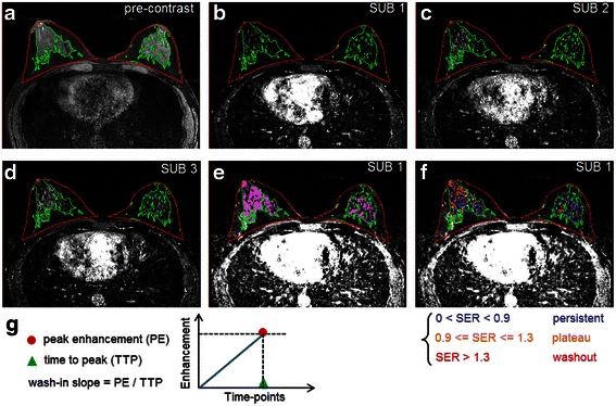
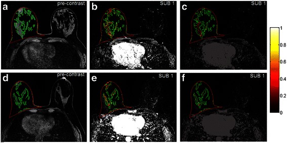
Under a Health Insurance Portability and Accountability Act compliant and Institutional Review Board-approved protocol, DCE-MRI scans of the contralateral breasts of 51 patients with cancer and 51 controls (matched by age and year of MRI) with biopsy-proven benign lesions were retrospectively analyzed. Applying fully automated computer algorithms on pre-contrast and multiple post-contrast MR sequences, two contrast enhancement kinetic variables, wash-in slope and signal enhancement ratio, were quantified from normal parenchyma of the contralateral breasts of both patients with cancer and controls. Conditional logistic regression was employed to assess association between these two measures and presence of breast cancer, with adjustment for other imaging factors including mammographic breast density and MRI background parenchymal enhancement (BPE). The area under the receiver operating characteristic curve (AUC) was used to assess the ability of the kinetic measures to distinguish patients with cancer from controls.
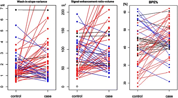
When both kinetic measures were included in conditional logistic regression analysis, the odds ratio for breast cancer was 1.7 (95 % CI 1.1, 2.8; p = 0.017) for wash-in slope variance and 3.5 (95 % CI 1.2, 9.9; p = 0.019) for signal enhancement ratio volume, respectively. These odds ratios were similar on respective univariate analysis, and remained significant after adjustment for menopausal status, family history, and mammographic density. While percent BPE was associated with an odds ratio of 3.1 (95 % CI 1.2, 7.9; p = 0.018), in multivariable analysis of the three measures, percent BPE was non-significant (p = 0.897) and the two kinetics measures remained significant. For the differentiation of patients with cancer and controls, the unadjusted AUC was 0.71 using a combination of the two measures, which significantly (p = 0.005) outperformed either measure alone (AUC = 0.65 for wash-in slope variance and 0.63 for signal enhancement ratio volume).
Kinetic measures of wash-in slope and signal enhancement ratio quantified from normal parenchyma in DCE-MRI are jointly associated with presence of breast cancer, even after adjustment for mammographic density and BPE.
在一项病例对照研究中,我们研究了从正常乳腺实质中量化的动态对比增强磁共振成像(DCE-MRI)对比增强动力学变量与乳腺癌存在情况的相关性。
根据符合《健康保险流通与责任法案》且经机构审查委员会批准的方案,对51例患有癌症的患者和51例对照者(根据年龄和MRI年份匹配)的对侧乳房进行DCE-MRI扫描,这些对照者经活检证实患有良性病变。在对比前和多个对比后的MR序列上应用全自动计算机算法,从癌症患者和对照者对侧乳房的正常实质中量化两个对比增强动力学变量,即流入斜率和信号增强率。采用条件逻辑回归评估这两个指标与乳腺癌存在情况之间的关联,并对其他影像因素进行调整,包括乳腺钼靶密度和MRI背景实质强化(BPE)。使用受试者操作特征曲线下面积(AUC)评估动力学指标区分癌症患者和对照者的能力。
当将这两个动力学指标纳入条件逻辑回归分析时,流入斜率方差的乳腺癌优势比为1.7(95%CI 1.1, 2.8;p = 0.017),信号增强率体积的优势比为3.5(95%CI 1.2, 9.9;p = 0.019)。在各自的单变量分析中,这些优势比相似,在对绝经状态、家族史和乳腺钼靶密度进行调整后仍具有显著性。虽然BPE百分比的优势比为3.1(95%CI 1.2, 7.9;p = 0.018),但在对这三个指标进行多变量分析时,BPE百分比无显著性(p = 0.897),而这两个动力学指标仍具有显著性。对于区分癌症患者和对照者,使用这两个指标组合的未调整AUC为0.71,显著优于单独使用任何一个指标(流入斜率方差的AUC = 0.65,信号增强率体积的AUC = 0.63;p = 0.005)。
即使在对乳腺钼靶密度和BPE进行调整后,DCE-MRI中从正常实质量化的流入斜率和信号增强率的动力学指标仍与乳腺癌的存在共同相关。