Departments of Radiology, Biomedical Informatics, and Bioengineering, University of Pittsburgh, 4200 Fifth Ave, Pittsburgh, PA, 15260, USA.
Magee-Womens Hospital of University of Pittsburgh Medical Center, 300 Halket St, Pittsburgh, PA, 15213, USA.
Sci Rep. 2017 May 18;7(1):2115. doi: 10.1038/s41598-017-02341-8.
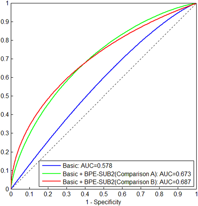
We investigated automated quantitative measures of background parenchymal enhancement (BPE) derived from an early versus delayed post-contrast sequence in breast dynamic contrast-enhanced magnetic resonance imaging (DCE-MRI) for association with breast cancer presence in a case-control study. DCE-MRIs were retrospectively analyzed for 51 cancer cases and 51 controls with biopsy-proven benign lesions, matched by age and year-of-MRI. BPE was quantified using fully-automated validated computer algorithms, separately from three sequential DCE-MRI post-contrast-subtracted sequences (SUB1, SUB2, and SUB3). The association of BPE computed from the three SUBs and other known factors with breast cancer were assessed in terms of odds ratio (OR) and area under the receiver operating characteristic curve (AUC). The OR of breast cancer for the percentage BPE measure (BPE%) quantified from SUB1 was 3.5 (95% Confidence Interval: 1.3, 9.8; p = 0.015) for 20% increments. Slightly lower and statistically significant ORs were also obtained for BPE quantified from SUB2 and SUB3. There was no significant difference (p > 0.2) in AUC for BPE quantified from the three post-contrast sequences and their combination. Our study showed that quantitative measures of BPE are associated with breast cancer presence and the association was similar across three breast DCE-MRI post-contrast sequences.
我们在一项病例对照研究中调查了乳腺动态对比增强磁共振成像(DCE-MRI)中早期与延迟对比后序列衍生的背景实质增强(BPE)的自动定量测量值与乳腺癌存在之间的关联。对 51 例癌症病例和 51 例经活检证实为良性病变的对照进行了回顾性 DCE-MRI 分析,这些对照按年龄和 MRI 年份匹配。使用完全自动化的验证计算机算法分别从三个连续的 DCE-MRI 对比后减去序列(SUB1、SUB2 和 SUB3)中定量 BPE。根据比值比(OR)和接受者操作特征曲线(ROC)下面积(AUC)评估从三个 SUB 计算的 BPE 和其他已知因素与乳腺癌的关联。从 SUB1 定量的 BPE%(BPE%)的乳腺癌 OR 为 3.5(95%置信区间:1.3, 9.8;p=0.015),BPE 增加 20%。从 SUB2 和 SUB3 定量的 BPE 也获得了略低但具有统计学意义的 OR。从三个对比后序列及其组合定量的 BPE 的 AUC 没有显著差异(p>0.2)。我们的研究表明,BPE 的定量测量值与乳腺癌的存在有关,并且在三个乳腺 DCE-MRI 对比后序列中,其相关性相似。