Llinás R, Sugimori M, Lin J W, Leopold P L, Brady S T
Department of Physiology and Biophysics, New York University Medical School, NY 10016.
Proc Natl Acad Sci U S A. 1989 Jul;86(14):5656-60. doi: 10.1073/pnas.86.14.5656.
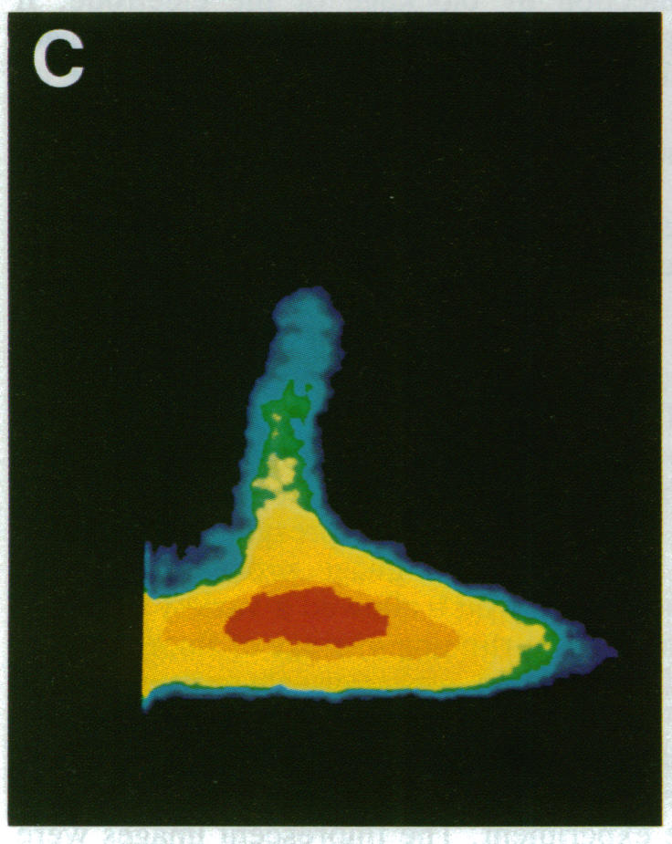
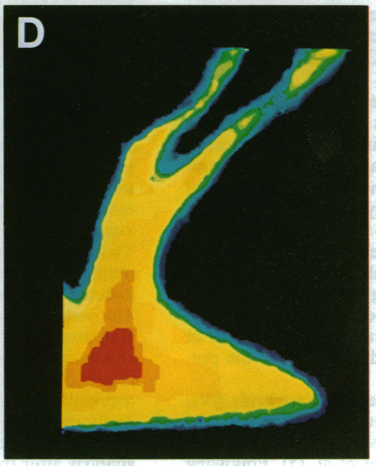
The question as to whether synaptic vesicles prepared from vertebrate brain can be transported to the active zones of the squid giant synapse was studied by using a combined optical and electrophysiological approach. In order to visualize the behavior of the vertebrate synaptic vesicles in situ, synaptic vesicles isolated from rat brain were labeled with a fluorescent dye (Texas red) and injected into the presynaptic terminal of the squid giant synapse. The pattern of fluorescence that would result from passive diffusion was determined by coinjection of an unconjugated fluorescent dye (fluorescein). The patterns obtained with fluorescent synaptic vesicles were strikingly different from that obtained by simple diffusion of fluorescein. Although the fluorescein diffused freely in both directions, the vesicles moved preferentially into the terminal--i.e., toward the release sites--at a rate of 0.5 microns/sec. The final distribution of the injected fluorescent synaptic vesicles displayed a discrete localization that suggested a distribution coincident with the active zones of the presynaptic terminal. Like fast axonal transport, but unlike fluorescein movements in the terminal, the vesicle movement was energy dependent, since the addition of 2,4-dinitrophenol blocked the redistribution of vesicles completely. In addition, reduction of extracellular calcium concentration reversibly blocked vesicular movement as well. In conclusion, mammalian synaptic vesicles retain the cytoplasmic surface components necessary for translocation, sorting, and targeting to the proper locations by the native machinery of the squid giant synapse.
通过光学和电生理相结合的方法,研究了从脊椎动物大脑制备的突触小泡是否能被运输到鱿鱼巨大突触的活性区。为了在原位观察脊椎动物突触小泡的行为,从大鼠大脑分离的突触小泡用荧光染料(德克萨斯红)标记,并注入鱿鱼巨大突触的突触前终末。通过共注射未结合的荧光染料(荧光素)来确定被动扩散产生的荧光模式。荧光突触小泡获得的模式与荧光素简单扩散获得的模式显著不同。虽然荧光素在两个方向上自由扩散,但小泡以0.5微米/秒的速度优先向终末移动,即向释放位点移动。注射的荧光突触小泡的最终分布显示出离散的定位,这表明其分布与突触前终末的活性区一致。与快速轴突运输一样,但与荧光素在终末的移动不同,小泡的移动是能量依赖的,因为添加2,4-二硝基苯酚完全阻断了小泡的重新分布。此外,细胞外钙浓度的降低也可逆地阻断了小泡的移动。总之,哺乳动物突触小泡保留了通过鱿鱼巨大突触的天然机制进行转运、分选和靶向到适当位置所需的细胞质表面成分。