Badoud Simon, Van De Ville Dimitri, Nicastro Nicolas, Garibotto Valentina, Burkhard Pierre R, Haller Sven
Neurology Division, Department of Clinical Neurosciences (NEUCLI), Geneva University Hospitals, Switzerland; Neurophysiology Unit, Department of Medicine, University of Fribourg (CH), Switzerland; Faculty of Medicine, University of Geneva, Switzerland.
Department of Imaging and Medical Informatics, University Hospitals of Geneva, Faculty of Medicine, University of Geneva, Switzerland; Institute of Bioengineering, Ecole Polytechnique Fédérale de Lausanne, Switzerland.
Neuroimage Clin. 2016 Jul 5;12:234-40. doi: 10.1016/j.nicl.2016.07.004. eCollection 2016.
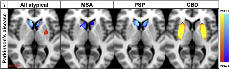
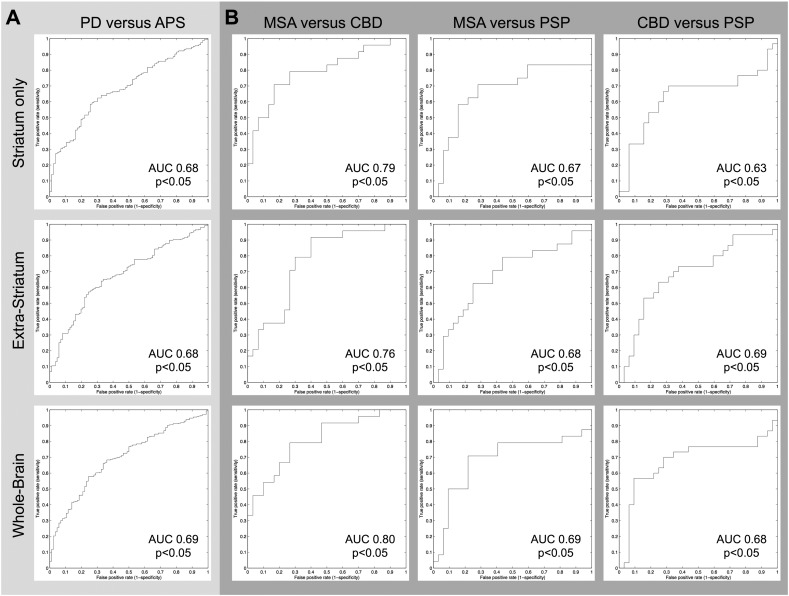
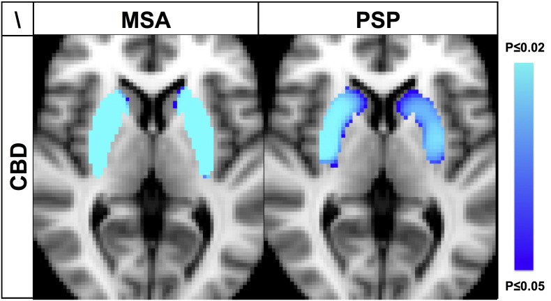
(123)I-ioflupane single photon emission computed tomography (SPECT) is a sensitive and well established imaging tool in Parkinson's disease (PD) and atypical parkinsonian syndromes (APS), yet a discrimination between PD and APS has been considered inconsistent at least based on visual inspection or simple region of interest analyses. We here reappraise this issue by applying advanced image analysis techniques to separate PD from the various APS. This study included 392 consecutive patients with degenerative parkinsonism undergoing (123)I-ioflupane SPECT at our institution over the last decade: 306 PD, 24 multiple system atrophy (MSA), 32 progressive supranuclear palsy (PSP) and 30 corticobasal degeneration (CBD) patients. Data analysis included voxel-wise univariate statistical parametric mapping and multivariate pattern recognition using linear discriminant classifiers. MSA and PSP showed less ioflupane uptake in the head of caudate nucleus relative to PD and CBD, yet there was no difference between MSA and PSP. CBD had higher uptake in both putamen relative to PD, MSA and PSP. Classification was significant for PD versus APS (AUC 0.69, p < 0.05) and between APS subtypes (MSA vs CBD AUC 0.80, p < 0.05; MSA vs PSP AUC 0.69 p < 0.05; CBD vs PSP AUC 0.69 p < 0.05). Both striatal and extra-striatal regions contain classification information, yet the combination of both regions does not significantly improve classification accuracy. PD, MSA, PSP and CBD have distinct patterns of dopaminergic depletion on (123)I-ioflupane SPECT. The high specificity of 84-90% for PD versus APS indicates that the classifier is particularly useful for confirming APS cases.
(123)I-碘氟潘单光子发射计算机断层扫描(SPECT)是帕金森病(PD)和非典型帕金森综合征(APS)中一种敏感且成熟的成像工具,但至少基于视觉检查或简单的感兴趣区域分析,PD和APS之间的鉴别一直被认为是不一致的。我们在此通过应用先进的图像分析技术来区分PD和各种APS,重新评估这个问题。本研究纳入了过去十年间在我们机构连续接受(123)I-碘氟潘SPECT检查的392例退行性帕金森综合征患者:306例PD患者、24例多系统萎缩(MSA)患者、32例进行性核上性麻痹(PSP)患者和30例皮质基底节变性(CBD)患者。数据分析包括体素水平的单变量统计参数映射和使用线性判别分类器的多变量模式识别。与PD和CBD相比,MSA和PSP在尾状核头部的碘氟潘摄取较少,但MSA和PSP之间没有差异。与PD、MSA和PSP相比,CBD在双侧壳核中的摄取较高。PD与APS之间(曲线下面积[AUC]为0.69,p<0.05)以及APS各亚型之间(MSA与CBD,AUC为0.80,p<0.05;MSA与PSP,AUC为0.69,p<0.05;CBD与PSP,AUC为0.69,p<0.05)的分类具有显著性。纹状体和纹状体以外的区域都包含分类信息,但两个区域的组合并没有显著提高分类准确性。PD、MSA、PSP和CBD在(123)I-碘氟潘SPECT上具有不同的多巴胺能缺失模式。PD与APS相比84-90%的高特异性表明该分类器对确诊APS病例特别有用。