• 文献检索
  • 文档翻译
  • 深度研究
  • 学术资讯
  • Suppr Zotero 插件Zotero 插件
  • 邀请有礼
  • 套餐&价格
  • 历史记录
应用&插件
Suppr Zotero 插件Zotero 插件浏览器插件Mac 客户端Windows 客户端微信小程序
定价
高级版会员购买积分包购买API积分包
服务
文献检索文档翻译深度研究API 文档MCP 服务
关于我们
关于 Suppr公司介绍联系我们用户协议隐私条款
关注我们

Suppr 超能文献

核心技术专利:CN118964589B侵权必究
粤ICP备2023148730 号-1Suppr @ 2026

文献检索

告别复杂PubMed语法,用中文像聊天一样搜索,搜遍4000万医学文献。AI智能推荐,让科研检索更轻松。

立即免费搜索

文件翻译

保留排版,准确专业,支持PDF/Word/PPT等文件格式,支持 12+语言互译。

免费翻译文档

深度研究

AI帮你快速写综述,25分钟生成高质量综述,智能提取关键信息,辅助科研写作。

立即免费体验

用于定量磁共振成像的T1和T2算法验证:通过独立于供应商的软件进行性能评估。

Validation of T1 and T2 algorithms for quantitative MRI: performance by a vendor-independent software.

作者信息

Bidhult Sebastian, Kantasis George, Aletras Anthony H, Arheden Håkan, Heiberg Einar, Hedström Erik

机构信息

Department of Clinical Sciences Lund, Clinical Physiology, Lund University, Skane University Hospital, Lund, Sweden.

Department of Biomedical Engineering, Faculty of Engineering, Lund University, Lund, Sweden.

出版信息

BMC Med Imaging. 2016 Aug 8;16(1):46. doi: 10.1186/s12880-016-0148-6.

DOI:10.1186/s12880-016-0148-6
PMID:27501697
原文链接:https://pmc.ncbi.nlm.nih.gov/articles/PMC4977731/
Abstract

BACKGROUND

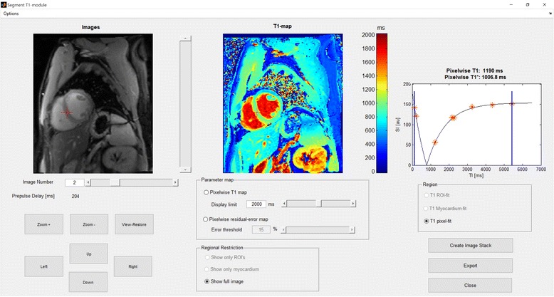
Determination of the relaxation time constants T1 and T2 with quantitative magnetic resonance imaging is increasingly used for both research and clinical practice. Recently, groups have been formed within the Society of Cardiovascular Magnetic Resonance to address issues with relaxometry. However, so far they have avoided specific recommendations on methodology due to lack of consensus and current evolving research. Standardised widely available software may simplify this process. The purpose of the current study was to develop and validate vendor-independent T1 and T2 mapping modules and implement those in the versatile and widespread software Segment, freely available for research and FDA approved for clinical applications.

RESULTS

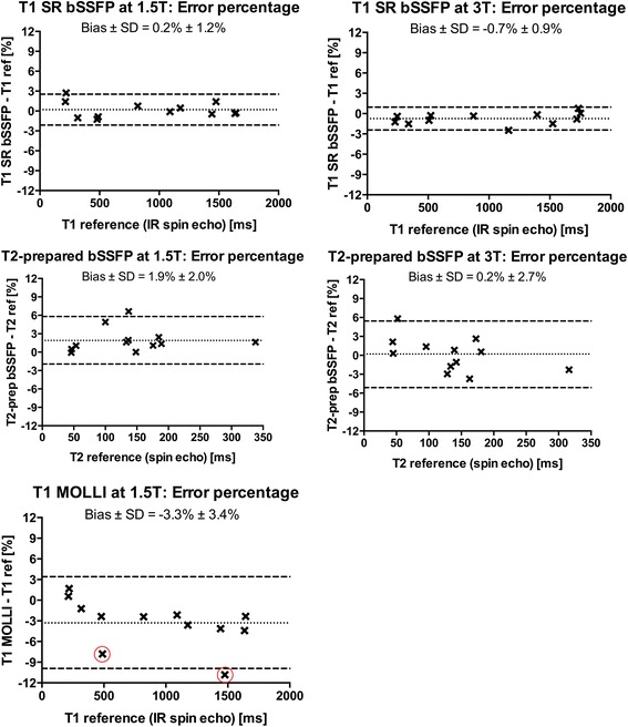
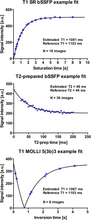
The T1 and T2 mapping modules were developed and validated in phantoms at 1.5 T and 3 T with reference standard values calculated from reference pulse sequences using the Nelder-Mead Simplex optimisation method. The proposed modules support current commonly available MRI pulse sequences and both 2- and 3-parameter curve fitting. Images acquired in patients using three major vendors showed vendor-independence. Bias and variability showed high agreement with T1 and T2 reference standards for T1 (range 214-1752 ms) and T2 (range 45-338 ms), respectively.

CONCLUSIONS

The developed and validated T1 and T2 mapping and quantification modules generated relaxation maps from current commonly used MRI sequences and multiple signal models. Patient applications showed usability for three major vendors.

摘要

背景

利用定量磁共振成像测定弛豫时间常数T1和T2在研究和临床实践中应用越来越广泛。最近,心血管磁共振学会内部已成立了多个小组来解决弛豫测量法相关问题。然而,由于缺乏共识以及当前研究仍在不断发展,到目前为止他们尚未就方法学给出具体建议。标准化的通用软件可能会简化这一过程。本研究的目的是开发并验证与设备供应商无关的T1和T2映射模块,并将其应用于通用且广泛使用的Segment软件中,该软件可免费用于研究,且已获得美国食品药品监督管理局的临床应用批准。

结果

T1和T2映射模块在1.5T和3T的体模中进行了开发和验证,通过使用Nelder-Mead单纯形优化方法从参考脉冲序列计算出参考标准值。所提出的模块支持当前常用的MRI脉冲序列以及二参数和三参数曲线拟合。使用三大主要设备供应商的设备在患者中采集的图像显示出与设备供应商无关。偏差和变异性分别与T1(范围为214 - 1752毫秒)和T2(范围为45 - 338毫秒)的T1和T2参考标准高度一致。

结论

所开发并经验证的T1和T2映射及量化模块可从当前常用的MRI序列和多种信号模型生成弛豫图。在患者中的应用表明该模块对三大主要设备供应商均适用。

https://cdn.ncbi.nlm.nih.gov/pmc/blobs/9d9e/4977731/a7ea39ebd694/12880_2016_148_Fig4_HTML.jpg
https://cdn.ncbi.nlm.nih.gov/pmc/blobs/9d9e/4977731/4ea02d0ce8bc/12880_2016_148_Fig1_HTML.jpg
https://cdn.ncbi.nlm.nih.gov/pmc/blobs/9d9e/4977731/05bdf1b50ed3/12880_2016_148_Fig2_HTML.jpg
https://cdn.ncbi.nlm.nih.gov/pmc/blobs/9d9e/4977731/9b96b71144e1/12880_2016_148_Fig3_HTML.jpg
https://cdn.ncbi.nlm.nih.gov/pmc/blobs/9d9e/4977731/a7ea39ebd694/12880_2016_148_Fig4_HTML.jpg

相似文献

1
Validation of T1 and T2 algorithms for quantitative MRI: performance by a vendor-independent software.用于定量磁共振成像的T1和T2算法验证:通过独立于供应商的软件进行性能评估。
BMC Med Imaging. 2016 Aug 8;16(1):46. doi: 10.1186/s12880-016-0148-6.
2
Magnetic resonance fingerprinting using echo-planar imaging: Joint quantification of T and T2∗ relaxation times.基于回波平面成像的磁共振指纹成像:T 和 T2*弛豫时间的联合定量。
Magn Reson Med. 2017 Nov;78(5):1724-1733. doi: 10.1002/mrm.26561. Epub 2016 Dec 16.
3
Pulmonary relaxometry with inversion recovery ultra-fast steady-state free precession at 1.5T.1.5T下采用反转恢复超快速稳态自由进动的肺部弛豫测量法。
Magn Reson Med. 2017 Jan;77(1):74-82. doi: 10.1002/mrm.26490. Epub 2016 Oct 19.
4
Establishing intra- and inter-vendor reproducibility of T relaxation time measurements with 3T MRI.建立 3T MRI 下 T 弛豫时间测量的内、外供应商重现性。
Magn Reson Med. 2019 Jan;81(1):454-465. doi: 10.1002/mrm.27421. Epub 2018 Aug 29.
5
Validation of a new T2* algorithm and its uncertainty value for cardiac and liver iron load determination from MRI magnitude images.一种用于从MRI幅度图像确定心脏和肝脏铁负荷的新型T2*算法及其不确定度值的验证。
Magn Reson Med. 2016 Apr;75(4):1717-29. doi: 10.1002/mrm.25767. Epub 2015 May 22.
6
An open-source software tool for the generation of relaxation time maps in magnetic resonance imaging.用于磁共振成像中弛豫时间图生成的开源软件工具。
BMC Med Imaging. 2010 Jul 30;10:16. doi: 10.1186/1471-2342-10-16.
7
How stable is quantitative MRI? - Assessment of intra- and inter-scanner-model reproducibility using identical acquisition sequences and data analysis programs.定量 MRI 的稳定性如何?——使用相同的采集序列和数据分析程序评估扫描仪内和扫描仪间模型的可重复性。
Neuroimage. 2020 Feb 15;207:116364. doi: 10.1016/j.neuroimage.2019.116364. Epub 2019 Nov 15.
8
Cross-vendor multiparametric mapping of the human brain using 3D-QALAS: A multicenter and multivendor study.使用 3D-QALAS 对人类大脑进行跨供应商的多参数映射:一项多中心和多供应商研究。
Magn Reson Med. 2024 May;91(5):1863-1875. doi: 10.1002/mrm.29939. Epub 2024 Jan 9.
9
Controlled saturation magnetization transfer for reproducible multivendor variable flip angle T and T mapping.用于可重复的多厂商可变翻转角T1和T2*映射的可控饱和磁化传递
Magn Reson Med. 2020 Jul;84(1):221-236. doi: 10.1002/mrm.28109. Epub 2019 Dec 17.
10
In vivo biochemical 7.0 Tesla magnetic resonance: preliminary results of dGEMRIC, zonal T2, and T2* mapping of articular cartilage.体内生物化学7.0特斯拉磁共振成像:关节软骨延迟钆增强磁共振成像、分区T2及T2*成像的初步结果
Invest Radiol. 2008 Sep;43(9):619-26. doi: 10.1097/RLI.0b013e31817e9122.

引用本文的文献

1
Differentiation of histological calcification classifications in breast cancer using ultrashort echo time and chemical shift-encoded imaging MRI.利用超短回波时间和化学位移编码成像MRI对乳腺癌组织学钙化分类进行鉴别
Front Oncol. 2024 Dec 17;14:1475090. doi: 10.3389/fonc.2024.1475090. eCollection 2024.
2
Phase-sensitive deep reconstruction method for rapid multiparametric MR fingerprinting and quantitative susceptibility mapping in the brain.用于脑部快速多参数磁共振指纹成像和定量磁化率映射的相敏深度重建方法。
Magn Reson Imaging. 2024 Jun;109:147-157. doi: 10.1016/j.mri.2024.03.023. Epub 2024 Mar 19.
3
Fetal growth restriction followed by very preterm birth is associated with smaller kidneys but preserved kidney function in adolescence.

本文引用的文献

1
Parallel simulations for QUAntifying RElaxation magnetic resonance constants (SQUAREMR): an example towards accurate MOLLI T1 measurements.用于量化弛豫磁共振常数的并行模拟(SQUAREMR):迈向准确MOLLI T1测量的一个示例
J Cardiovasc Magn Reson. 2015 Nov 26;17:104. doi: 10.1186/s12968-015-0206-1.
2
Prostate cancer discrimination in the peripheral zone with a reduced field-of-view T(2)-mapping MRI sequence.使用缩小视野的T(2) 映射MRI序列在外周区鉴别前列腺癌。
Magn Reson Imaging. 2015 Jun;33(5):525-30. doi: 10.1016/j.mri.2015.02.006. Epub 2015 Feb 14.
3
Quantitative T1 and T2 mapping in recurrent glioblastomas under bevacizumab: earlier detection of tumor progression compared to conventional MRI.
胎儿生长受限继而出现极早产与青春期时肾脏较小但肾功能正常有关。
Pediatr Nephrol. 2023 Jun;38(6):1855-1866. doi: 10.1007/s00467-022-05785-x. Epub 2022 Nov 21.
4
Long-Term Tri-Modal In Vivo Tracking of Engrafted Cartilage-Derived Stem/Progenitor Cells Based on Upconversion Nanoparticles.基于上转换纳米粒子的移植软骨源性干细胞/祖细胞的长期三模态体内追踪。
Biomolecules. 2021 Jun 29;11(7):958. doi: 10.3390/biom11070958.
5
Characterization of acute TLR-7 agonist-induced hemorrhagic myocarditis in mice by multiparametric quantitative cardiac magnetic resonance imaging.多参数定量心脏磁共振成像对小鼠急性 TLR-7 激动剂诱导的出血性心肌炎的特征分析。
Dis Model Mech. 2019 Aug 16;12(8):dmm040725. doi: 10.1242/dmm.040725.
6
Myocardial tissue characterization in patients with hereditary gelsolin (AGel) amyloidosis using novel cardiovascular magnetic resonance techniques.使用新型心血管磁共振技术对遗传性凝溶胶蛋白(AGel)淀粉样变性患者的心肌组织特征进行研究。
Int J Cardiovasc Imaging. 2019 Feb;35(2):351-358. doi: 10.1007/s10554-019-01570-4. Epub 2019 Mar 8.
贝伐单抗治疗复发性胶质母细胞瘤的定量T1和T2映射:与传统MRI相比,可更早检测肿瘤进展
Neuroradiology. 2015 Jan;57(1):11-20. doi: 10.1007/s00234-014-1445-9. Epub 2014 Oct 7.
4
Optimized saturation recovery protocols for T1-mapping in the heart: influence of sampling strategies on precision.心脏T1映射的优化饱和恢复协议:采样策略对精度的影响。
J Cardiovasc Magn Reson. 2014 Sep 4;16(1):55. doi: 10.1186/s12968-014-0055-3.
5
Improved quantitative myocardial T mapping: Impact of the fitting model.改进的定量心肌T值映射:拟合模型的影响
Magn Reson Med. 2015 Jul;74(1):93-105. doi: 10.1002/mrm.25377. Epub 2014 Aug 7.
6
T1-mapping in the heart: accuracy and precision.心脏 T1-mapping:准确性和精密度。
J Cardiovasc Magn Reson. 2014 Jan 4;16(1):2. doi: 10.1186/1532-429X-16-2.
7
Myocardial T1 mapping and extracellular volume quantification: a Society for Cardiovascular Magnetic Resonance (SCMR) and CMR Working Group of the European Society of Cardiology consensus statement.心肌 T1 映射和细胞外容积定量:心血管磁共振学会 (SCMR) 和欧洲心脏病学会 CMR 工作组的共识声明。
J Cardiovasc Magn Reson. 2013 Oct 14;15(1):92. doi: 10.1186/1532-429X-15-92.
8
Saturation recovery single-shot acquisition (SASHA) for myocardial T(1) mapping.用于心肌T(1) 图谱分析的饱和恢复单次采集(SASHA)
Magn Reson Med. 2014 Jun;71(6):2082-95. doi: 10.1002/mrm.24878. Epub 2013 Jul 23.
9
Standardized image interpretation and post processing in cardiovascular magnetic resonance: Society for Cardiovascular Magnetic Resonance (SCMR) board of trustees task force on standardized post processing.心血管磁共振标准化图像解读和后处理:心血管磁共振学会(SCMR)董事会后处理标准化工作组。
J Cardiovasc Magn Reson. 2013 May 1;15(1):35. doi: 10.1186/1532-429X-15-35.
10
Phase-sensitive inversion recovery for myocardial T1 mapping with motion correction and parametric fitting.相位敏感反转恢复心肌 T1 映射与运动校正和参数拟合。
Magn Reson Med. 2013 May;69(5):1408-20. doi: 10.1002/mrm.24385. Epub 2012 Jun 26.