Lim Seung-Jae, Park Yoon-Soo
Department of Orthopaedic Surgery, Samsung Medical Center, Sungkyunkwan University School of Medicine, Seoul, Korea.
Hip Pelvis. 2015 Sep;27(3):125-34. doi: 10.5371/hp.2015.27.3.125. Epub 2015 Sep 30.
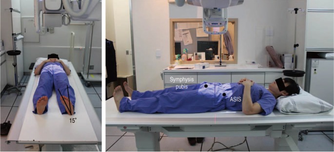
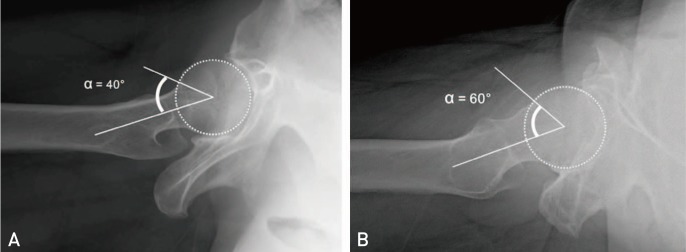
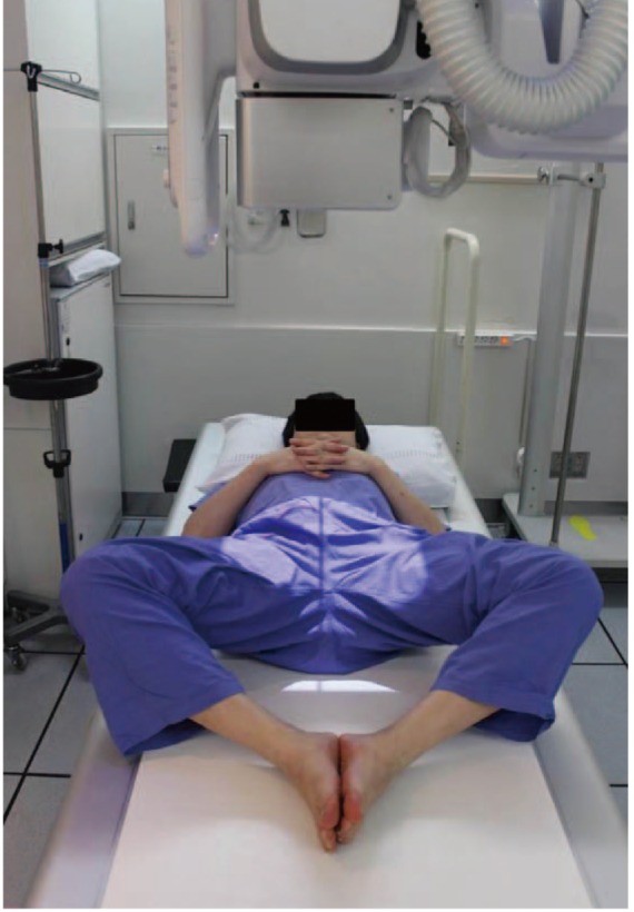
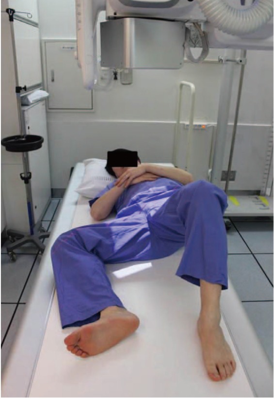
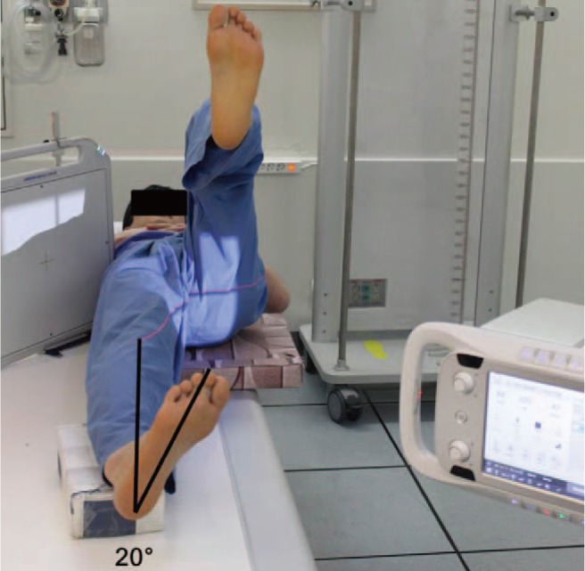
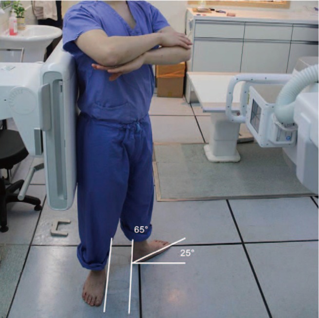
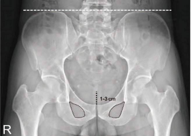
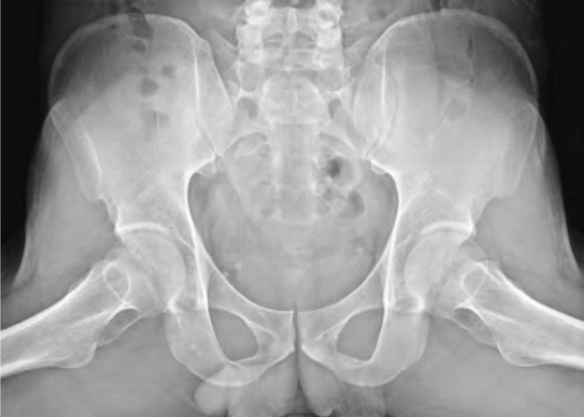
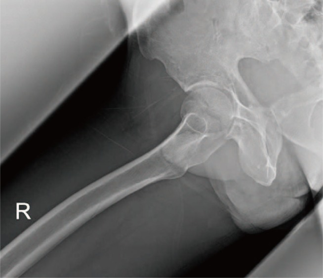
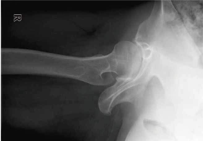
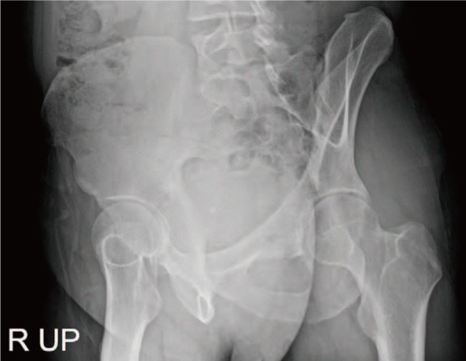
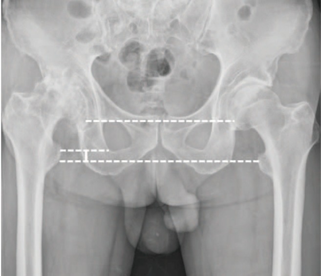
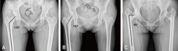
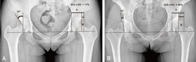
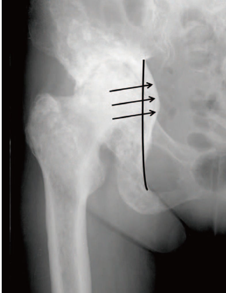
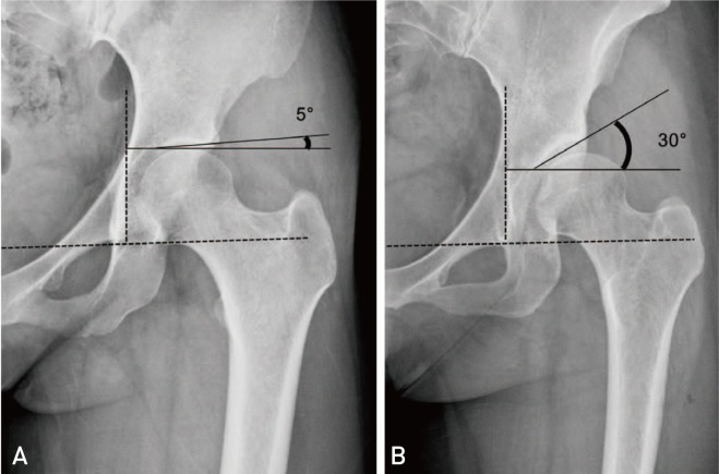
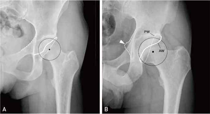
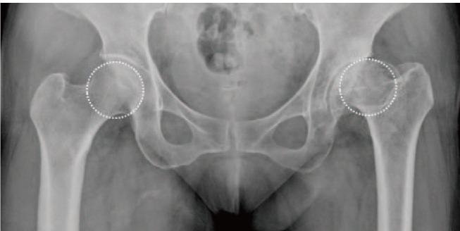
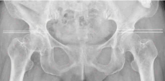
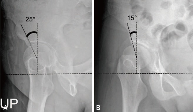
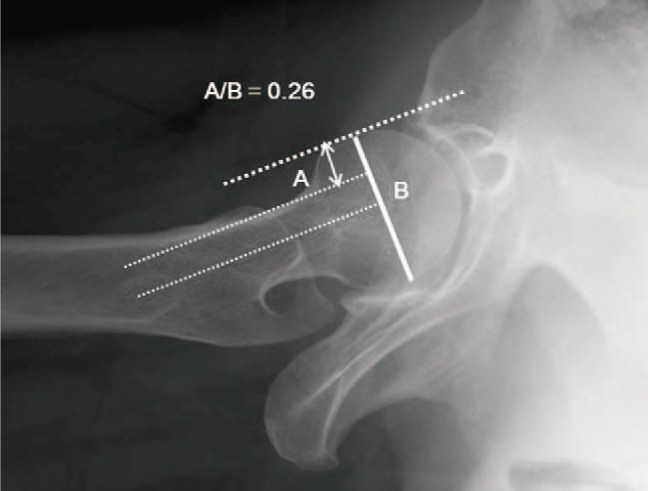
Plain radiographic examination is a fundamental approach to the diagnosis and treatment decision-making of the hip. A thorough understanding of standard radiographic techniques, radiographic anatomy, and disease patterns affecting the hip can be helpful in improving diagnostic accuracy. This article reviews the standard protocols used to obtain radiographic projections of the hip and addresses specific signs and various radiographic measurements used to adequately and reliably recognize structural diseases of the hip.
普通X线检查是髋关节诊断和治疗决策的基本方法。全面了解标准X线技术、X线解剖结构以及影响髋关节的疾病模式,有助于提高诊断准确性。本文回顾了用于获取髋关节X线投影的标准方案,并阐述了用于充分且可靠地识别髋关节结构疾病的特定体征和各种X线测量方法。