Yi Seung Rim, Lee Min Ho, Yang Bo Kyu, Ahn Young Joon, Kwon Jieun, Im Se Hyuk, Lee Ye Hyun
Department of Orthopedic Surgery, National Police Hospital, Seoul, Korea.
Hip Pelvis. 2015 Dec;27(4):265-72. doi: 10.5371/hp.2015.27.4.265. Epub 2015 Dec 30.
To assess the progression of clinical symptoms and disease course of calcific tendinitis in the hip region according to types of calcification.
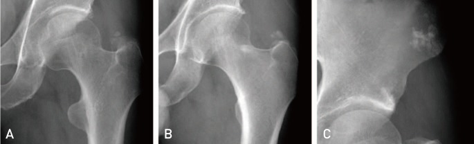
Among patients with the hip pain, 28 patients (21 males and 7 females; mean age 51 years, range 32-74 years) showing calcified lesions in simple radiography without other possible sources of pain were analyzed retrospectively. Twelve patients displayed a symptom duration of less than three weeks (acute; average=1±0.9 week) and 16 displayed greater than three weeks (chronic; average=21.0±19.5 weeks). Lesions were classified as nodular (11, 39.3%), nodular-fragmented (13, 46.4%), or amorphous (4, 14.3%). Initial symptoms, progression of clinical features, radiological findings and prognosis were investigated and analyzed according to calcification type.
In 15 patients (53.6%), lesions were located superior to the great trochanter. On average, the acute group was younger (44.58 vs. 55.44 years, P=0.006), suffered more (mean pain Numeric Rating Scale [NRS], 6.3 vs. 3.8; P<0.001), and recovered more (difference between initial and follow-up NRS, 5.1 vs. 2.63; <<0.001) than the chronic group. The mean length of initial lesions was longer in the acute group than the chronic group (15.8 vs. 9.1 mm, P=0.008). When compared to patients with distinctive margins (15, 53.6%), those with nondistinctive margins showed better improvement (difference between initial and follow-up NRS, 4.7 vs. 2.8; P=0.01) and more significant decrease in lesion size (difference between initial and follow-up length, 10.8 vs. 2.6 mm; P=0.003).
Calcific tendinitis occurring in the hip area displayed a variety of characteristics. Although complaining of more severe pain in the initial phase, patients with acute pain or calcific lesions with nondistinctive margins showed better symptom improvement when compared to their counterparts.
根据钙化类型评估髋部钙化性肌腱炎的临床症状进展和病程。
回顾性分析28例髋部疼痛患者(男21例,女7例;平均年龄51岁,范围32 - 74岁),这些患者在X线平片上显示有钙化病变,且无其他可能的疼痛来源。12例患者症状持续时间小于3周(急性;平均 = 1±0.9周),16例患者症状持续时间大于3周(慢性;平均 = 21.0±19.5周)。病变分为结节状(11例,39.3%)、结节 - 碎片状(13例,46.4%)或无定形(4例,14.3%)。根据钙化类型对初始症状、临床特征进展、影像学表现和预后进行调查分析。
15例患者(53.6%)的病变位于大转子上方。平均而言,急性组患者比慢性组患者更年轻(44.58岁对55.44岁,P = 0.006),疼痛更严重(平均疼痛数字评分量表[NRS],6.3对3.8;P < 0.001),恢复情况更好(初始与随访NRS差值,5.1对2.63;P << 0.001)。急性组初始病变的平均长度比慢性组长(15.8对9.1 mm,P = 0.008)。与边缘清晰的患者(15例,53.6%)相比,边缘不清晰的患者症状改善更好(初始与随访NRS差值,4.7对2.8;P = 0.01),病变大小减小更显著(初始与随访长度差值,10.8对2.6 mm;P = 0.003)。
髋部发生的钙化性肌腱炎表现出多种特征。尽管在初始阶段疼痛更严重,但与慢性疼痛患者或边缘清晰的钙化病变患者相比,急性疼痛患者或边缘不清晰的钙化病变患者症状改善更好。