Newey C R, Sarwal A, Hantus S
Department of Neurology, University of Missouri, 5 Hospital Drive, CE 540, Columbia, MO 65211, USA.
Neurology and Critical Care, Wake Forest University School of Medicine, Reynolds M, Medical Center Boulevard, Winston-Salem, NC 27157, USA.
Autoimmune Dis. 2016;2016:9450452. doi: 10.1155/2016/9450452. Epub 2016 Jul 31.
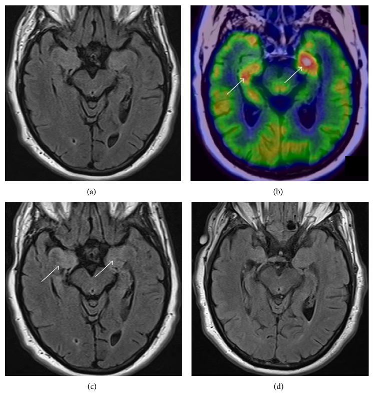
Introduction. Autoimmune encephalitis (AE) is a clinically challenging diagnosis with nonspecific neurological symptoms. Prompt diagnosis is important and often relies on neuroimaging. We present a case series of AE highlighting the importance of an early [(18)F]-fluoro-deoxy-glucose positron emission tomography (FDG-PET) scan. Methods. Retrospective review of seven consecutive cases of autoimmune encephalitis. Results. All patients had both magnetic resonance imaging (MRI) and FDG-PET scans. Initial clinical presentations included altered mental status and/or new onset seizures. Six cases had serum voltage-gated potassium channel (VGKC) antibody and one had serum N-methyl-D-aspartate (NMDA) antibody. MRI of brain showed mesial temporal lobe hyperintensity in five cases of VGKC. The other two patients with VGKC or NMDA AE had restiform body hyperintensity on MRI brain or a normal MRI, respectively. Mesial temporal lobe hypermetabolism was noted in three cases on FDG-PET, despite initial unremarkable MRI. Malignancy workup was negative in all patients. Conclusion. A high index of suspicion for AE should be maintained in patients presenting with cognitive symptoms, seizures, and limbic changes on neuroimaging. In cases with normal initial brain MRI, FDG-PET can be positive. Additionally, extralimbic hyperintensity on MRI may also be observed.
引言。自身免疫性脑炎(AE)是一种临床诊断具有挑战性的疾病,其神经症状不具特异性。及时诊断很重要,且往往依赖于神经影像学检查。我们展示了一组自身免疫性脑炎病例系列,强调了早期[(18)F] - 氟脱氧葡萄糖正电子发射断层扫描(FDG - PET)的重要性。方法。对连续7例自身免疫性脑炎病例进行回顾性分析。结果。所有患者均进行了磁共振成像(MRI)和FDG - PET扫描。初始临床表现包括精神状态改变和/或新发癫痫。6例患者血清电压门控钾通道(VGKC)抗体阳性,1例患者血清N - 甲基 - D - 天冬氨酸(NMDA)抗体阳性。5例VGKC抗体阳性患者的脑部MRI显示内侧颞叶高信号。另外2例VGKC或NMDA抗体阳性的自身免疫性脑炎患者,MRI分别显示绳状体高信号或MRI正常。尽管初始MRI无明显异常,但3例患者的FDG - PET显示内侧颞叶代谢增高。所有患者的恶性肿瘤检查均为阴性。结论。对于出现认知症状、癫痫发作且神经影像学检查有边缘叶改变的患者,应高度怀疑自身免疫性脑炎。在初始脑部MRI正常的病例中,FDG - PET可能呈阳性。此外,MRI上也可能观察到边缘叶外高信号。