Bissig Christin, Rochin Leila, van Niel Guillaume
Institut Curie, Paris Sciences et Lettres Research University, UMR144, Centre de Recherche, 26 rue d'ULM, Paris F-75231, France.
Centre National de la Recherche Scientifique, UMR144, Paris F-75248, France.
Int J Mol Sci. 2016 Aug 31;17(9):1438. doi: 10.3390/ijms17091438.
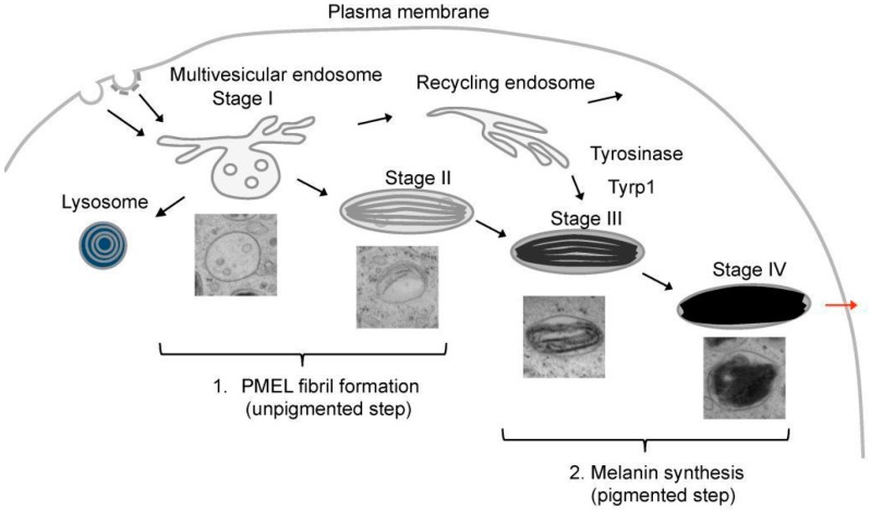
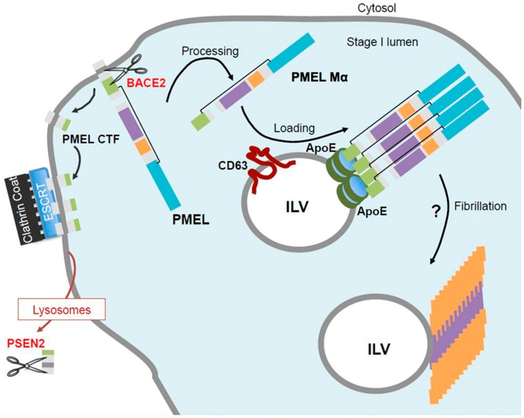
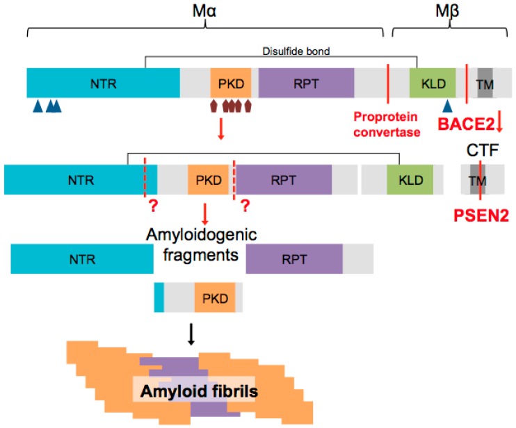
In pigment cells, melanin synthesis takes place in specialized organelles, called melanosomes. The biogenesis and maturation of melanosomes is initiated by an unpigmented step that takes place prior to the initiation of melanin synthesis and leads to the formation of luminal fibrils deriving from the pigment cell-specific pre-melanosomal protein (PMEL). In the lumen of melanosomes, PMEL fibrils optimize sequestration and condensation of the pigment melanin. Interestingly, PMEL fibrils have been described to adopt a typical amyloid-like structure. In contrast to pathological amyloids often associated with neurodegenerative diseases, PMEL fibrils represent an emergent category of physiological amyloids due to their beneficial cellular functions. The formation of PMEL fibrils within melanosomes is tightly regulated by diverse mechanisms, such as PMEL traffic, cleavage and sorting. These mechanisms revealed increasing analogies between the formation of physiological PMEL fibrils and pathological amyloid fibrils. In this review we summarize the known mechanisms of PMEL fibrillation and discuss how the recent understanding of physiological PMEL amyloid formation may help to shed light on processes involved in pathological amyloid formation.
在色素细胞中,黑色素合成发生在称为黑素小体的特殊细胞器中。黑素小体的生物发生和成熟始于黑色素合成开始之前的一个无色素步骤,该步骤导致形成源自色素细胞特异性前黑素小体蛋白(PMEL)的腔内原纤维。在黑素小体的腔内,PMEL原纤维优化了色素黑色素的隔离和浓缩。有趣的是,PMEL原纤维已被描述为具有典型的淀粉样蛋白样结构。与通常与神经退行性疾病相关的病理性淀粉样蛋白不同,PMEL原纤维由于其有益的细胞功能而代表了一类新兴的生理性淀粉样蛋白。黑素小体内PMEL原纤维的形成受到多种机制的严格调控,如PMEL的运输、切割和分选。这些机制揭示了生理性PMEL原纤维形成与病理性淀粉样蛋白原纤维形成之间越来越多的相似之处。在这篇综述中,我们总结了已知的PMEL纤维化机制,并讨论了对生理性PMEL淀粉样蛋白形成的最新理解如何有助于阐明病理性淀粉样蛋白形成所涉及的过程。