Qian Yunzhu, Chen Hanbang, Xu Yang, Yang Jianxin, Zhou Xuefeng, Zhang Feimin, Gu Ning
Jiangsu Key Laboratory of Oral Diseases, Nanjing Medical University, Nanjing; Center of Stomatology, The Second Affiliated Hospital of Soochow University, Suzhou.
Jiangsu Key Laboratory of Oral Diseases, Nanjing Medical University, Nanjing.
Int J Nanomedicine. 2016 Aug 25;11:4157-71. doi: 10.2147/IJN.S110577. eCollection 2016.
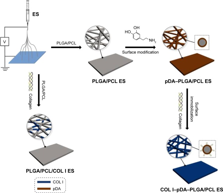
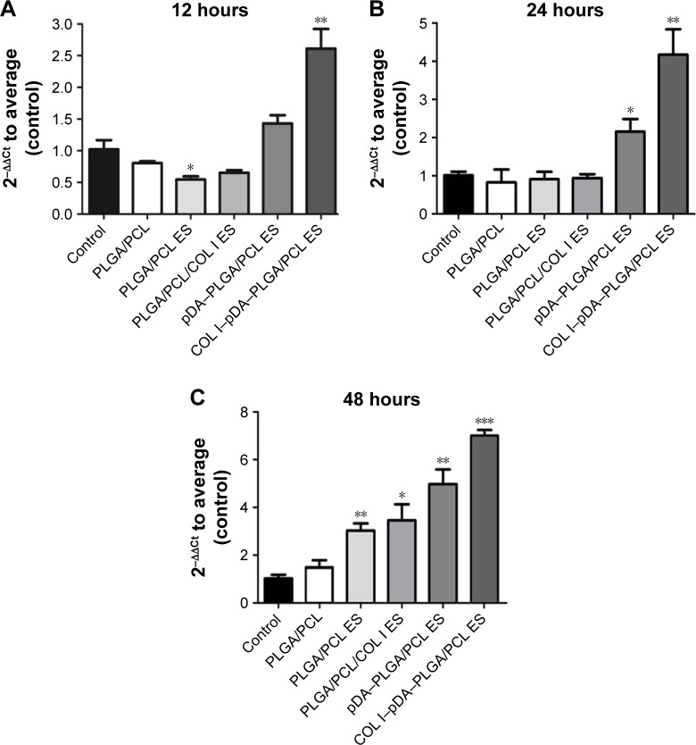
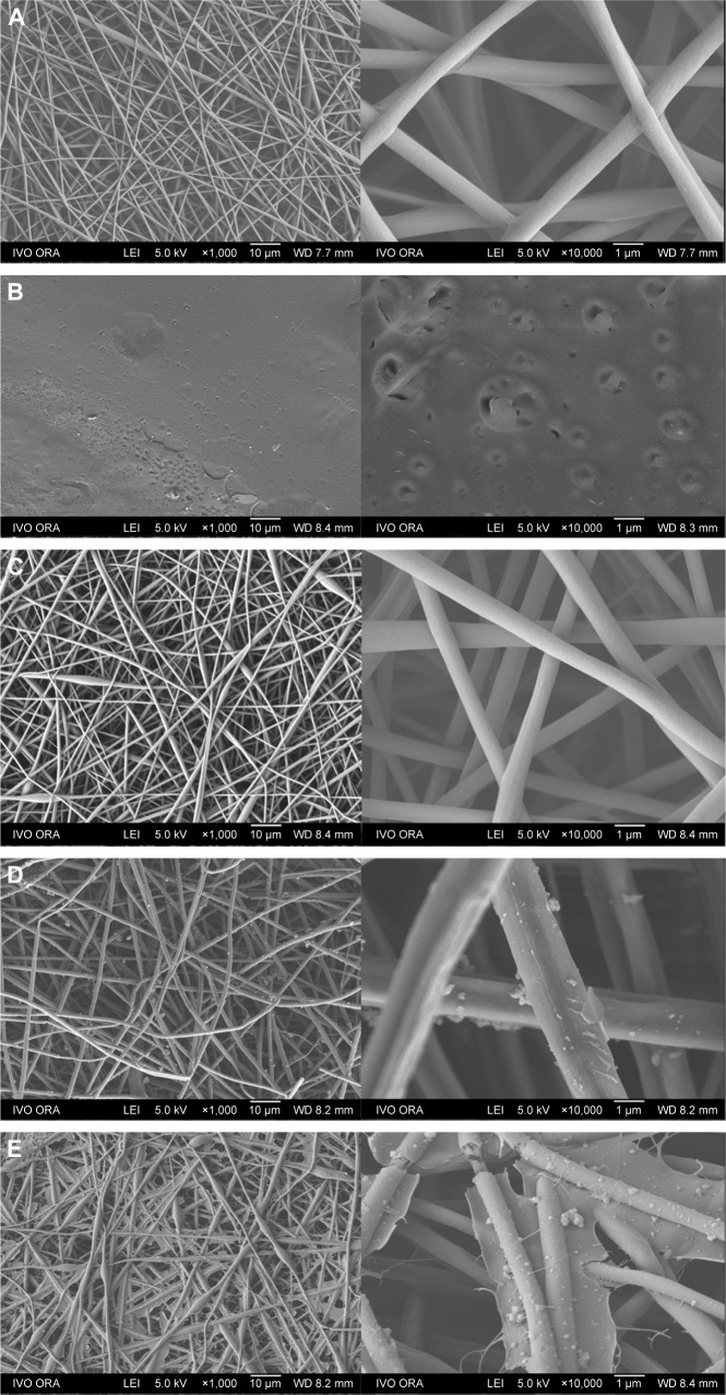
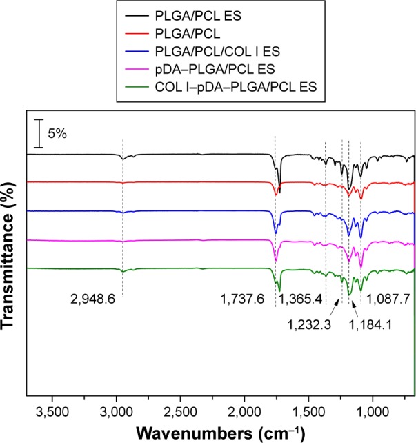
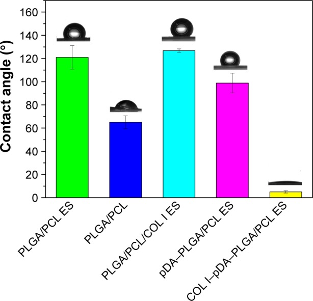
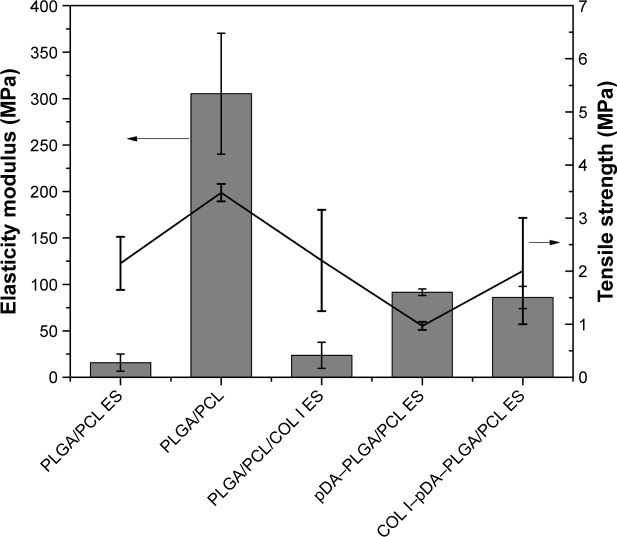
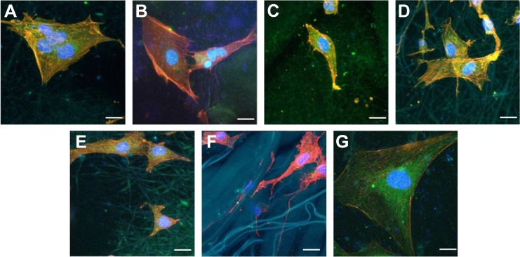
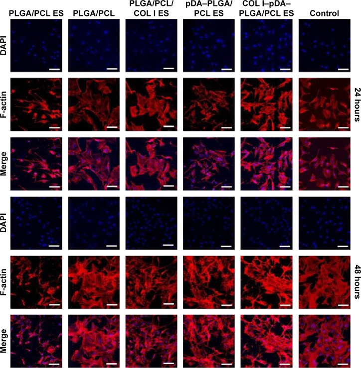
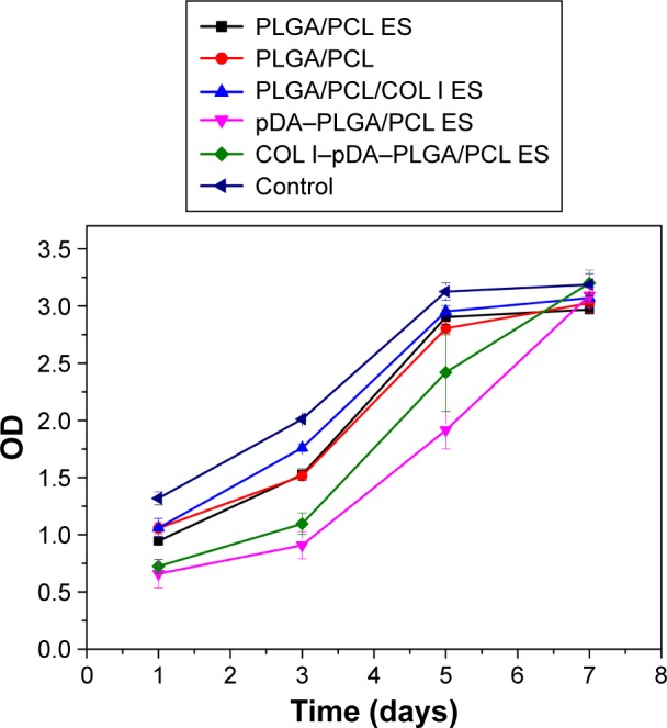
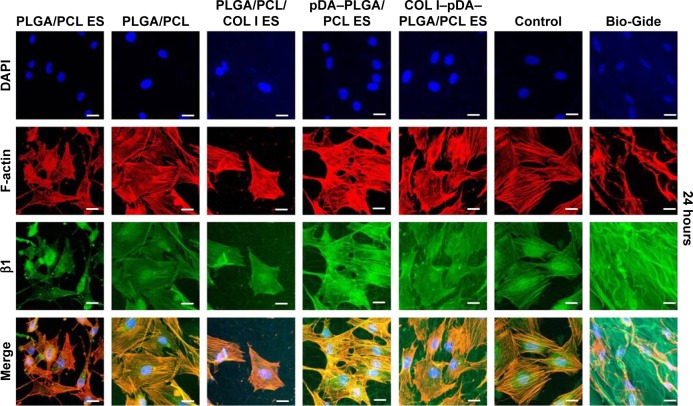
Constructing biomimetic structure and incorporating bioactive molecules is an effective strategy to achieve a more favorable cell response. To explore the effect of electrospinning (ES) nanofibrous architecture and collagen I (COL I)-incorporated modification on tuning osteoblast response, a resorbable membrane composed of poly(lactic-co-glycolic acid)/poly(caprolactone) (PLGA/PCL; 7:3 w/w) was developed via ES. COL I was blended into PLGA/PCL solution to prepare composite ES membrane. Notably, relatively better cell response was delivered by the bioactive ES-based membrane which was fabricated by modification of 3,4-dihydroxyphenylalanine and COL I. After investigation by field emission scanning electron microscopy, Fourier transform infrared spectroscopy, contact angle measurement, and mechanical test, polyporous three-dimensional nanofibrous structure with low tensile force and the successful integration of COL I was obtained by the ES method. Compared with traditional PLGA/PCL membrane, the surface hydrophilicity of collagen-incorporated membranes was largely enhanced. The behavior of mouse preosteoblast MC3T3-E1 cell infiltration and proliferation on membranes was studied at 24 and 48 hours. The negative control was fabricated by solvent casting. Evaluation of cell adhesion and morphology demonstrated that all the ES membranes were more favorable for promoting the cell adhesion and spreading than the casting membrane. Cell Counting Kit-8 assays revealed that biomimetic architecture, surface topography, and bioactive properties of membranes were favorable for cell growth. Analysis of β1 integrin expression level by immunofluorescence indicated that such biomimetic architecture, especially COL I-grafted surface, plays a key role in cell adhesion and proliferation. The real-time polymerase chain reaction suggested that both surface topography and bioactive properties could facilitate the cell adhesion. The combined effect of biomimetic architecture with enhanced surface activity by 3,4-dihydroxyphenylalanine-assisted modification and COL I incorporation of PLGA/PCL electrospun membranes could successfully fill osteogenic defects and allow for better cell proliferation and differentiation.
构建仿生结构并掺入生物活性分子是实现更有利细胞反应的有效策略。为了探究静电纺丝(ES)纳米纤维结构和掺入I型胶原蛋白(COL I)的修饰对调节成骨细胞反应的影响,通过ES制备了一种由聚(乳酸-共-乙醇酸)/聚己内酯(PLGA/PCL;7:3 w/w)组成的可吸收膜。将COL I混入PLGA/PCL溶液中以制备复合ES膜。值得注意的是,通过3,4-二羟基苯丙氨酸和COL I修饰制备的基于生物活性ES的膜表现出相对更好的细胞反应。经过场发射扫描电子显微镜、傅里叶变换红外光谱、接触角测量和力学测试研究,通过ES方法获得了具有低拉伸力的多孔三维纳米纤维结构以及COL I的成功整合。与传统的PLGA/PCL膜相比,掺入胶原蛋白的膜的表面亲水性大大增强。在24小时和48小时研究了小鼠前成骨细胞MC3T3-E1在膜上的浸润和增殖行为。阴性对照通过溶剂浇铸制备。细胞黏附及形态评估表明,所有ES膜在促进细胞黏附和铺展方面都比浇铸膜更有利。细胞计数试剂盒-8检测显示,膜的仿生结构、表面形貌和生物活性特性有利于细胞生长。通过免疫荧光分析β1整合素表达水平表明,这种仿生结构,尤其是COL I接枝表面,在细胞黏附和增殖中起关键作用。实时聚合酶链反应表明,表面形貌和生物活性特性都可以促进细胞黏附。通过3,4-二羟基苯丙氨酸辅助修饰和掺入COL I增强表面活性的PLGA/PCL静电纺丝膜的仿生结构的联合作用可以成功填充成骨缺损,并实现更好的细胞增殖和分化。