• 文献检索
  • 文档翻译
  • 深度研究
  • 学术资讯
  • Suppr Zotero 插件Zotero 插件
  • 邀请有礼
  • 套餐&价格
  • 历史记录
应用&插件
Suppr Zotero 插件Zotero 插件浏览器插件Mac 客户端Windows 客户端微信小程序
定价
高级版会员购买积分包购买API积分包
服务
文献检索文档翻译深度研究API 文档MCP 服务
关于我们
关于 Suppr公司介绍联系我们用户协议隐私条款
关注我们

Suppr 超能文献

核心技术专利:CN118964589B侵权必究
粤ICP备2023148730 号-1Suppr @ 2026

文献检索

告别复杂PubMed语法,用中文像聊天一样搜索,搜遍4000万医学文献。AI智能推荐,让科研检索更轻松。

立即免费搜索

文件翻译

保留排版,准确专业,支持PDF/Word/PPT等文件格式,支持 12+语言互译。

免费翻译文档

深度研究

AI帮你快速写综述,25分钟生成高质量综述,智能提取关键信息,辅助科研写作。

立即免费体验

通过眼前节光学相干断层扫描和活体共聚焦显微镜测量角膜和角膜缘上皮厚度。

Measurement of corneal and limbal epithelial thickness by anterior segment optical coherence tomography and in vivo confocal microscopy.

作者信息

Le Qihua, Chen Yan, Yang Yujing, Xu Jianjiang

机构信息

Department of Ophthalmology, Eye & ENT Hospital of Fudan University, No. 83 Fenyang Road, Shanghai, 200031, China.

Research Center, Eye & ENT Hospital of Fudan University, Shanghai, 200031, China.

出版信息

BMC Ophthalmol. 2016 Sep 20;16(1):163. doi: 10.1186/s12886-016-0342-x.

DOI:10.1186/s12886-016-0342-x
PMID:27645227
原文链接:https://pmc.ncbi.nlm.nih.gov/articles/PMC5029042/
Abstract

BACKGROUND

To compare corneal epithelial thickness (CET) and limbal epithelial thickness (LET) measured by anterior segment optical coherence tomography (AS-OCT) and in vivo confocal microscope (IVCM) in normal subjects, and evaluate the consistency between them.

METHODS

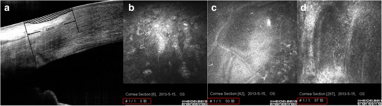
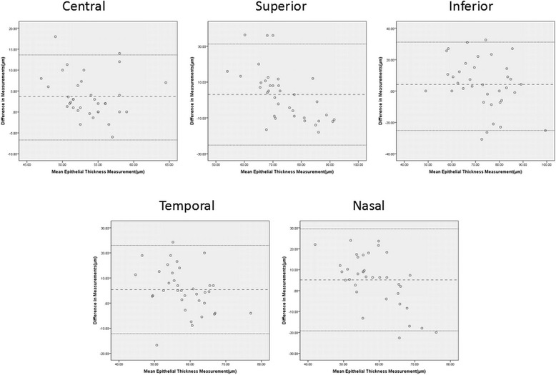
Thirty-eight normal subjects (17 men and 21 women) were enrolled in this study. AS-OCT was performed at central cornea and the superior, inferior, nasal and temporal limbus. Then followed by IVCM examination performed at the same location. Agreement was analyzed by mean difference (AS-OCT minus IVCM), 95 % limits of agreement (LoA) (1.96 standard deviation of the difference), and Bland-Altman analysis.

RESULTS

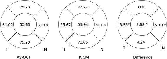
The average CET measured by AS-OCT and IVCM was 55.6 ± 4.0 μm and 51.9 ± 4.9 μm respectively. The value measured by IVCM was significantly lower than that measured by AS-OCT (P = 0.015). The average LET values tested by AS-OCT were 10.3 and 10.9 % less at nasal and temporal quadrant (nasal: P = 0.019, temporal: P = 0.003), and were similar as those measured by IVCM at superior and inferior quadrant. In subjects older than 40 years, CET and LET values measured by AS-OCT were significantly higher than those by IVCM. Such differences were not found in subjects ≤ 40 years old.

CONCLUSIONS

CET values measured by IVCM are lower than those by AS-OCT, while LET values measured by two devices have good agreement. These two techniques have their own advantages in measuring epithelial thickness and are mutually complementary.

摘要

背景

比较正常受试者中通过眼前节光学相干断层扫描(AS-OCT)和活体共聚焦显微镜(IVCM)测量的角膜上皮厚度(CET)和角膜缘上皮厚度(LET),并评估两者之间的一致性。

方法

本研究纳入38名正常受试者(17名男性和21名女性)。在中央角膜以及上方、下方、鼻侧和颞侧角膜缘进行AS-OCT检查。然后在相同位置进行IVCM检查。通过平均差异(AS-OCT减去IVCM)、95%一致性界限(LoA)(差异的1.96倍标准差)和Bland-Altman分析来分析一致性。

结果

AS-OCT和IVCM测量的平均CET分别为55.6±4.0μm和51.9±4.9μm。IVCM测量的值显著低于AS-OCT测量的值(P = 0.015)。AS-OCT检测的鼻侧和颞侧象限的平均LET值分别低10.3%和10.9%(鼻侧:P = 0.019,颞侧:P = 0.003),与IVCM在上方和下方象限测量的值相似。在40岁以上的受试者中,AS-OCT测量的CET和LET值显著高于IVCM测量的值。在≤40岁的受试者中未发现此类差异。

结论

IVCM测量的CET值低于AS-OCT测量的值,而两种设备测量的LET值具有良好的一致性。这两种技术在测量上皮厚度方面各有优势,且相互补充。

https://cdn.ncbi.nlm.nih.gov/pmc/blobs/eaf9/5029042/0dab5328bca2/12886_2016_342_Fig3_HTML.jpg
https://cdn.ncbi.nlm.nih.gov/pmc/blobs/eaf9/5029042/cbf0ef5a81c3/12886_2016_342_Fig1_HTML.jpg
https://cdn.ncbi.nlm.nih.gov/pmc/blobs/eaf9/5029042/1e7c44618ea6/12886_2016_342_Fig2_HTML.jpg
https://cdn.ncbi.nlm.nih.gov/pmc/blobs/eaf9/5029042/0dab5328bca2/12886_2016_342_Fig3_HTML.jpg
https://cdn.ncbi.nlm.nih.gov/pmc/blobs/eaf9/5029042/cbf0ef5a81c3/12886_2016_342_Fig1_HTML.jpg
https://cdn.ncbi.nlm.nih.gov/pmc/blobs/eaf9/5029042/1e7c44618ea6/12886_2016_342_Fig2_HTML.jpg
https://cdn.ncbi.nlm.nih.gov/pmc/blobs/eaf9/5029042/0dab5328bca2/12886_2016_342_Fig3_HTML.jpg

相似文献

1
Measurement of corneal and limbal epithelial thickness by anterior segment optical coherence tomography and in vivo confocal microscopy.通过眼前节光学相干断层扫描和活体共聚焦显微镜测量角膜和角膜缘上皮厚度。
BMC Ophthalmol. 2016 Sep 20;16(1):163. doi: 10.1186/s12886-016-0342-x.
2
Corneal Epithelial Thickness Measured Using Anterior Segment Optical Coherence Tomography as a Diagnostic Parameter for Limbal Stem Cell Deficiency.利用眼前节光学相干断层扫描测量角膜上皮厚度作为诊断角膜缘干细胞缺陷的参数。
Am J Ophthalmol. 2020 Aug;216:132-139. doi: 10.1016/j.ajo.2020.04.006. Epub 2020 Apr 10.
3
In vivo confocal microscopy and anterior segment optical coherence tomography analysis of the cornea in nephropathic cystinosis.肾病性胱氨酸病患者角膜的体内共聚焦显微镜检查和眼前节光学相干断层扫描分析
Ophthalmology. 2009 May;116(5):870-6. doi: 10.1016/j.ophtha.2008.11.021.
4
Photophobia and corneal crystal density in nephropathic cystinosis: an in vivo confocal microscopy and anterior-segment optical coherence tomography study.肾病性胱氨酸病中的畏光与角膜晶体密度:一项共聚焦显微镜活体检查及眼前节光学相干断层扫描研究
Invest Ophthalmol Vis Sci. 2015 May;56(5):3218-25. doi: 10.1167/iovs.15-16499.
5
Association of Changes in Thickness of Limbal Epithelial and Stroma with Corneal Scars Detected by High-Resolution Anterior Segment Optic Coherence Tomography.通过高分辨率眼前节光学相干断层扫描检测到的角膜缘上皮和基质厚度变化与角膜瘢痕的相关性
Klin Monbl Augenheilkd. 2024 Jun;241(6):744-750. doi: 10.1055/a-1842-2683. Epub 2022 May 3.
6
Age-related changes in human corneal epithelial thickness measured with anterior segment optical coherence tomography.使用眼前节光学相干断层扫描测量的人角膜上皮厚度的年龄相关性变化。
Invest Ophthalmol Vis Sci. 2014 Jul 22;55(8):5032-8. doi: 10.1167/iovs.13-13831.
7
En-face optical coherence tomography as a novel tool for exploring the ocular surface: a pilot comparative study to conventional B-scans and in vivo confocal microscopy.眼前节光学相干断层扫描作为一种探索眼表的新工具:与传统 B 扫描和活体共聚焦显微镜的初步对比研究。
Ocul Surf. 2014 Oct;12(4):285-306. doi: 10.1016/j.jtos.2014.02.006. Epub 2014 Jul 11.
8
Correlation between the existence of the palisades of Vogt and limbal epithelial thickness in limbal stem cell deficiency.Vogt栅栏的存在与角膜缘干细胞缺乏症中角膜缘上皮厚度之间的相关性。
Clin Exp Ophthalmol. 2017 Apr;45(3):224-231. doi: 10.1111/ceo.12832. Epub 2016 Oct 11.
9
Diagnosis of limbal stem cell deficiency based on corneal epithelial thickness measured on anterior segment optical coherence tomography.基于眼前节光学相干断层扫描测量的角膜上皮厚度诊断角膜缘干细胞缺乏症。
Indian J Ophthalmol. 2017 Nov;65(11):1120-1126. doi: 10.4103/ijo.IJO_218_17.
10
Assessment of corneal and limbal epithelial thickness by spectral-domain optical coherence tomography in brachycephalic and non-brachycephalic dogs.通过光谱域光学相干断层扫描评估短头犬和非短头犬的角膜和角膜缘上皮厚度
Vet Ophthalmol. 2023 Apr;26 Suppl 1:89-97. doi: 10.1111/vop.13016. Epub 2022 Jul 29.

引用本文的文献

1
Corneal epithelial thickness profile at different corneal zones of normal eyes in a population sample from northern Iran: A cross-sectional study.伊朗北部人群样本中正常眼睛不同角膜区域的角膜上皮厚度分布:一项横断面研究。
Oman J Ophthalmol. 2025 Jun 24;18(2):144-149. doi: 10.4103/ojo.ojo_236_23. eCollection 2025 May-Aug.
2
Corneal epithelium is altered in keratoconus and forme fruste keratoconus.圆锥角膜和亚临床圆锥角膜的角膜上皮会发生改变。
Sci Rep. 2025 Jul 1;15(1):22203. doi: 10.1038/s41598-025-07663-6.
3
The Corneal Epithelial Thickness Profile in a Healthy Saudi Population.

本文引用的文献

1
Comparison of Corneal Epithelial Thickness Measurement Between Fourier-Domain OCT and Very High-Frequency Digital Ultrasound.傅里叶域光学相干断层扫描与甚高频数字超声测量角膜上皮厚度的比较
J Refract Surg. 2015 Jul;31(7):438-45. doi: 10.3928/1081597X-20150623-01.
2
Epithelial Thinning in Limbal Stem Cell Deficiency.角膜缘干细胞缺乏症中的上皮变薄
Am J Ophthalmol. 2015 Oct;160(4):669-77.e4. doi: 10.1016/j.ajo.2015.06.029. Epub 2015 Jul 7.
3
Corneal epithelial thickness mapping using Fourier-domain optical coherence tomography for detection of form fruste keratoconus.
沙特健康人群的角膜上皮厚度分布
Cureus. 2024 Oct 9;16(10):e71135. doi: 10.7759/cureus.71135. eCollection 2024 Oct.
4
Repeatability of Corneal Epithelial Thickness Map Using Anterior-Segment Optical Coherence Tomography in Normal and Corneal Disease Patients.眼前节光学相干断层扫描测量正常人和角膜病患者角膜上皮厚度图的可重复性
Korean J Ophthalmol. 2024 Dec;38(6):496-506. doi: 10.3341/kjo.2024.0115. Epub 2024 Oct 22.
5
Three-Month Comprehensive Outcomes of Topography-Guided LASIK Versus Keratorefractive Lenticule Extraction (KLEx): A Prospective Contralateral Study.地形图引导的准分子激光原位角膜磨镶术与角膜屈光透镜切除术(KLEx)的三个月综合结果:一项前瞻性对侧研究。
Ophthalmol Ther. 2024 Aug;13(8):2265-2284. doi: 10.1007/s40123-024-00987-y. Epub 2024 Jul 1.
6
Corneal epithelial thickness mapping.角膜上皮厚度地形图绘制
Indian J Ophthalmol. 2024 Feb 1;72(2):155-156. doi: 10.4103/IJO.IJO_21_24. Epub 2024 Jan 25.
7
Corneal Epithelial Thickness Mapping: A Major Review.角膜上皮厚度地形图:一项主要综述。
J Ophthalmol. 2024 Jan 2;2024:6674747. doi: 10.1155/2024/6674747. eCollection 2024.
8
Diagnostic Accuracy of Corneal and Epithelial Thickness Map Parameters to Detect Keratoconus and Suspect Keratoconus.用于检测圆锥角膜和疑似圆锥角膜的角膜及上皮厚度图参数的诊断准确性
J Ophthalmol. 2023 Oct 6;2023:6677932. doi: 10.1155/2023/6677932. eCollection 2023.
9
Role of corneal epithelial mapping, Corvis biomechanical index, and artificial intelligence-based tomographic biomechanical index in diagnosing spectrum of keratoconus.角膜上皮地形图、Corvis生物力学指数及基于人工智能的断层生物力学指数在圆锥角膜诊断中的作用
Oman J Ophthalmol. 2023 May 26;16(2):276-280. doi: 10.4103/ojo.ojo_336_22. eCollection 2023 May-Aug.
10
Understanding Corneal Epithelial Thickness Mapping.了解角膜上皮厚度测绘。
Middle East Afr J Ophthalmol. 2023 May 25;29(3):147-155. doi: 10.4103/meajo.meajo_207_22. eCollection 2022 Jul-Sep.
使用傅里叶域光学相干断层扫描进行角膜上皮厚度测绘以检测forme fruste圆锥角膜。
J Cataract Refract Surg. 2015 Apr;41(4):812-20. doi: 10.1016/j.jcrs.2014.06.043.
4
Corneal epithelial thickness map in long-term soft contact lenses wearers.长期佩戴软性隐形眼镜者的角膜上皮厚度图。
Optom Vis Sci. 2014 Dec;91(12):1455-61. doi: 10.1097/OPX.0000000000000410.
5
Age-related changes in human corneal epithelial thickness measured with anterior segment optical coherence tomography.使用眼前节光学相干断层扫描测量的人角膜上皮厚度的年龄相关性变化。
Invest Ophthalmol Vis Sci. 2014 Jul 22;55(8):5032-8. doi: 10.1167/iovs.13-13831.
6
Tear film measurement by optical reflectometry technique.泪膜测量的光学反射测量技术。
J Biomed Opt. 2014 Feb;19(2):027001. doi: 10.1117/1.JBO.19.2.027001.
7
Age-related changes and diseases of the ocular surface and cornea.眼表和角膜的年龄相关性变化和疾病。
Invest Ophthalmol Vis Sci. 2013 Dec 13;54(14):ORSF48-53. doi: 10.1167/iovs.13-12840.
8
Anatomic alterations in aging and age-related diseases of the eye.眼的衰老和与年龄相关疾病的解剖学改变。
Invest Ophthalmol Vis Sci. 2013 Dec 13;54(14):ORSF23-7. doi: 10.1167/iovs.13-12711.
9
The corneal epithelial basement membrane: structure, function, and disease.角膜上皮基底膜:结构、功能与疾病。
Invest Ophthalmol Vis Sci. 2013 Sep 27;54(9):6390-400. doi: 10.1167/iovs.13-12547.
10
Measurement of tear film thickness using ultrahigh-resolution optical coherence tomography.使用超高分辨率光相干断层扫描测量泪膜厚度。
Invest Ophthalmol Vis Sci. 2013 Aug 15;54(8):5578-83. doi: 10.1167/iovs.13-11920.