Shah Karan M, Orton Peter, Mani Nick, Wilkinson Jeremy Mark, Gartland Alison
Department of Oncology and Metabolism, The University of Sheffield, Beech Hill Rd, Sheffield S10 2RX, United Kingdom.
J Orthop Res. 2017 Aug;35(8):1716-1723. doi: 10.1002/jor.23449. Epub 2016 Oct 14.
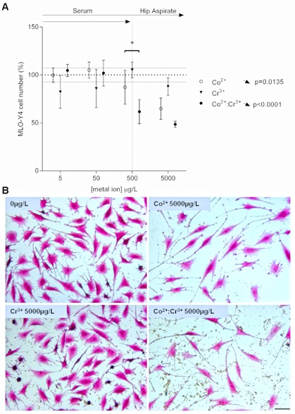
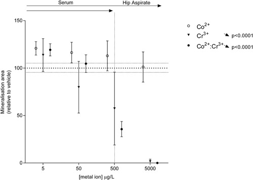
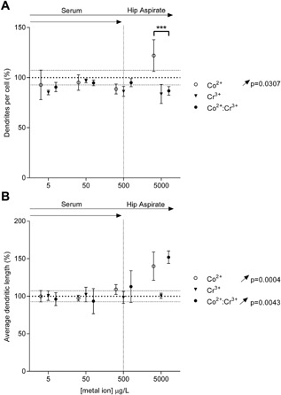
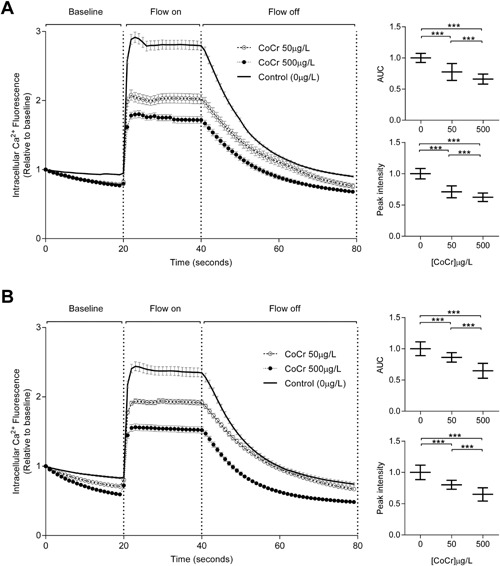
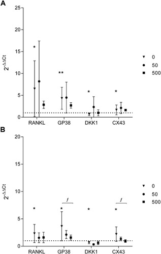
The effects of metal ion exposure on osteocytes, the most abundant cell type in bone and responsible for coordinating bone remodeling, remain unclear. However, several studies have previously shown that exposure to cobalt (Co ) and chromium (Cr ), at concentrations equivalent to those found clinically, affect osteoblast and osteoclast survival and function. In this study, we tested the hypothesis that metal ions would similarly impair the normal physiology of osteocytes. The survival, dendritic morphology, and response to fluid shear stress of the mature osteocyte-like cell-line MLO-Y4 following exposure to clinically relevant concentrations and combinations of Co and Cr ions were measured in 2D-culture. Exposure of MLO-Y4 cells to metal ions reduced cell number, increased dendrites per cell and increased dendrite length. We found that combinations of metal ions had a greater effect than the individual ions alone, and that Co had a predominate effect on changes to cell numbers and dendrites. Combined metal ion exposure blunted the responses of the MLO-Y4 cells to fluid shear stress, including reducing the intracellular calcium responses and modulation of genes for the osteocyte markers Cx43 and Gp38, and the signaling molecules RANKL and Dkk-1. Finally, we demonstrated that in the late osteoblasts/early osteocytes cell line MLO-A5 that Co exposure had no effect on mineralization, but Cr treatment inhibited mineralization in a dose-dependent manner, without affecting cell viability. Taken together, these data indicate that metal exposure can directly affect osteocyte physiology, with potential implications for bone health including osseointegration of cementless components, and periprosthetic bone remodeling. © 2016 The Authors. Journal of Orthopaedic Research Published by Wiley Periodicals, Inc. on behalf of Orthopaedic Research Society. J Orthop Res 35:1716-1723, 2017.
金属离子暴露对骨细胞(骨骼中最丰富的细胞类型,负责协调骨重塑)的影响尚不清楚。然而,此前多项研究表明,暴露于临床等效浓度的钴(Co)和铬(Cr)会影响成骨细胞和破骨细胞的存活及功能。在本研究中,我们检验了金属离子会同样损害骨细胞正常生理功能的假设。在二维培养中,测量了成熟的类骨细胞系MLO - Y4暴露于临床相关浓度及Co和Cr离子组合后的存活率、树突形态以及对流体剪切应力的反应。MLO - Y4细胞暴露于金属离子后,细胞数量减少,每个细胞的树突增多且树突长度增加。我们发现金属离子组合比单独的离子具有更大的影响,并且Co对细胞数量和树突的变化起主要作用。联合金属离子暴露减弱了MLO - Y4细胞对流体剪切应力的反应,包括降低细胞内钙反应以及调节骨细胞标志物Cx43和Gp38、信号分子RANKL和Dkk - 1的基因。最后,我们证明在晚期成骨细胞/早期骨细胞系MLO - A5中,Co暴露对矿化没有影响,但Cr处理以剂量依赖方式抑制矿化,且不影响细胞活力。综上所述,这些数据表明金属暴露可直接影响骨细胞生理功能,对骨骼健康包括非骨水泥部件的骨整合和假体周围骨重塑具有潜在影响。© 2016作者。《矫形外科研究杂志》由Wiley Periodicals, Inc.代表矫形外科研究协会出版。《矫形外科研究》35:1716 - 1723, 2017。