Shah Karan M, Dunning Mark J, Gartland Alison, Wilkinson J Mark
The Mellanby Centre for Musculoskeletal Research, Department of Oncology and Metabolism, The University of Sheffield, Beech Hill Rd, Sheffield S10 2RX, UK.
Sheffield Bioinformatics Core, The University of Sheffield, 385a Glossop Rd, Sheffield S10 2HQ, UK.
Int J Mol Sci. 2021 May 14;22(10):5225. doi: 10.3390/ijms22105225.
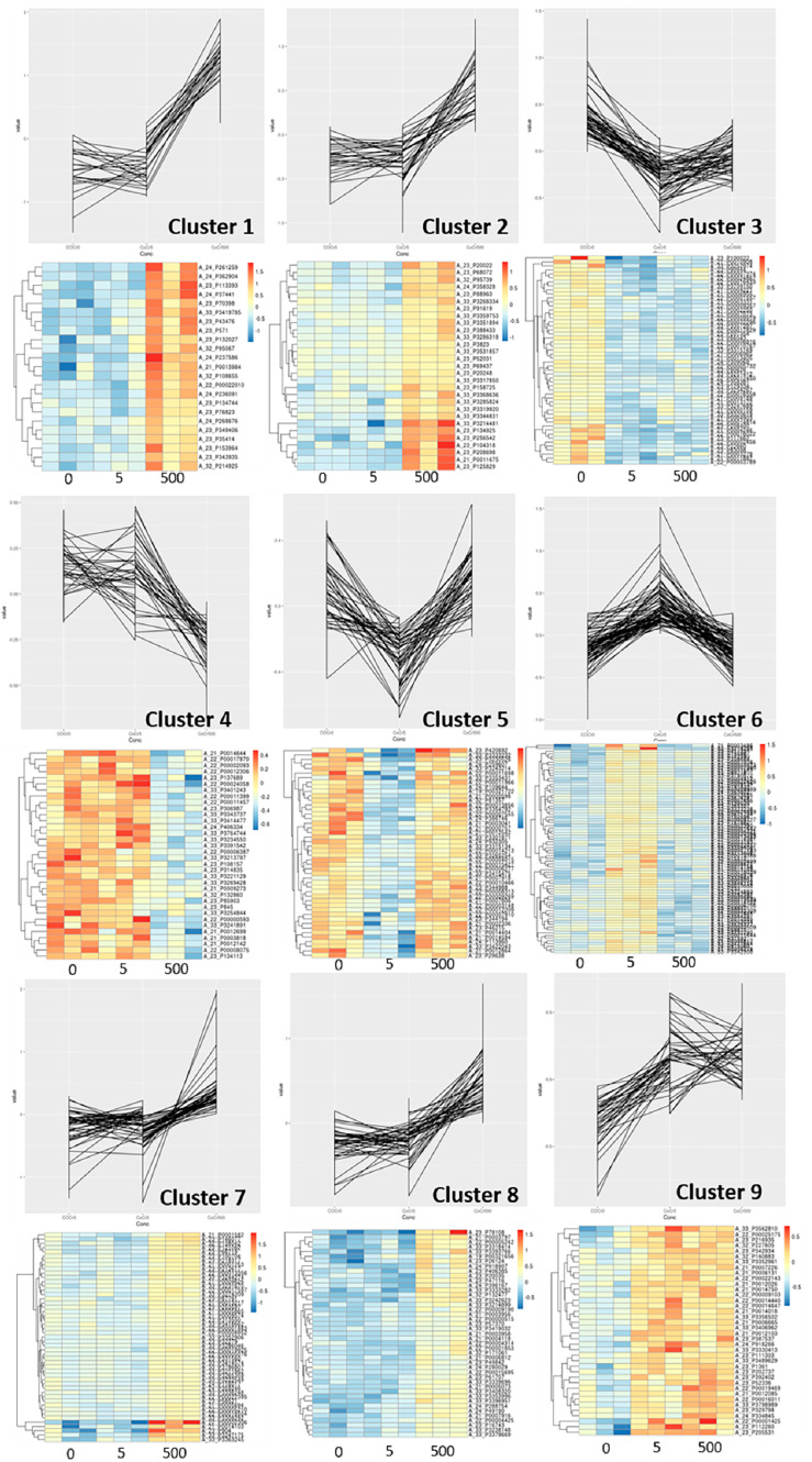
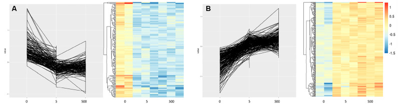
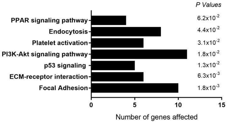
Systemic cobalt (Co) and chromium (Cr) concentrations may be elevated in patients with metal joint replacement prostheses. Several studies have highlighted the detrimental effects of this exposure on bone cells in vitro, but the underlying mechanisms remain unclear. In this study, we use whole-genome microarrays to comprehensively assess gene expression in primary human osteoblasts, osteoclast precursors and mature resorbing osteoclasts following exposure to clinically relevant circulating versus local periprosthetic tissue concentrations of Co and Cr ions and CoCr nanoparticles. We also describe the gene expression response in osteoblasts on routinely used prosthesis surfaces in the presence of metal exposure. Our results suggest that systemic levels of metal exposure have no effect on osteoblasts, and primarily inhibit osteoclast differentiation and function via altering the focal adhesion and extracellular matrix interaction pathways. In contrast, periprosthetic levels of metal exposure inhibit both osteoblast and osteoclast activity by altering HIF-1α signaling and endocytic/cytoskeletal genes respectively, as well as increasing inflammatory signaling with mechanistic implications for adverse reactions to metal debris. Furthermore, we identify gene clusters and KEGG pathways for which the expression correlates with increasing Co:Cr concentrations, and has the potential to serve as early markers of metal toxicity. Finally, our study provides a molecular basis for the improved clinical outcomes for hydroxyapatite-coated prostheses that elicit a pro-survival osteogenic gene signature compared to grit-blasted and plasma-sprayed titanium-coated surfaces in the presence of metal exposure.
金属关节置换假体患者的全身钴(Co)和铬(Cr)浓度可能会升高。多项研究强调了这种暴露在体外对骨细胞的有害影响,但其潜在机制仍不清楚。在本研究中,我们使用全基因组微阵列来全面评估原代人成骨细胞、破骨细胞前体和成熟吸收性破骨细胞在暴露于临床相关的循环与假体周围局部组织浓度的Co和Cr离子以及CoCr纳米颗粒后的基因表达。我们还描述了在存在金属暴露的情况下,常规使用的假体表面上成骨细胞的基因表达反应。我们结果表明,全身金属暴露水平对成骨细胞没有影响,并且主要通过改变粘着斑和细胞外基质相互作用途径来抑制破骨细胞分化和功能。相比之下,假体周围金属暴露水平分别通过改变HIF-1α信号通路和内吞/细胞骨架基因来抑制成骨细胞和破骨细胞活性,以及增加炎症信号通路,这对金属碎屑的不良反应具有机制上的影响。此外,我们确定了基因簇和KEGG通路,其表达与Co:Cr浓度升高相关,并且有可能作为金属毒性的早期标志物。最后,我们的研究为羟基磷灰石涂层假体在存在金属暴露的情况下与喷砂和等离子喷涂钛涂层表面相比引发促生存成骨基因特征从而改善临床结果提供了分子基础。