Martínez-Pinilla Eva, Ordóñez Cristina, Del Valle Eva, Navarro Ana, Tolivia Jorge
Departamento de Morfología y Biología Celular, Facultad de Medicina, Instituto de Neurociencias del Principado de Asturias, Universidad de Oviedo Oviedo, Spain.
Front Aging Neurosci. 2016 Sep 13;8:213. doi: 10.3389/fnagi.2016.00213. eCollection 2016.
Learning processes or language development are only some of the cognitive functions that differ qualitatively between men and women. Gender differences in the brain structure seem to be behind these variations. Indeed, this sexual dimorphism at neuroanatomical level is accompanied unequivocally by differences in the way that aging and neurodegenerative diseases affect men and women brains.
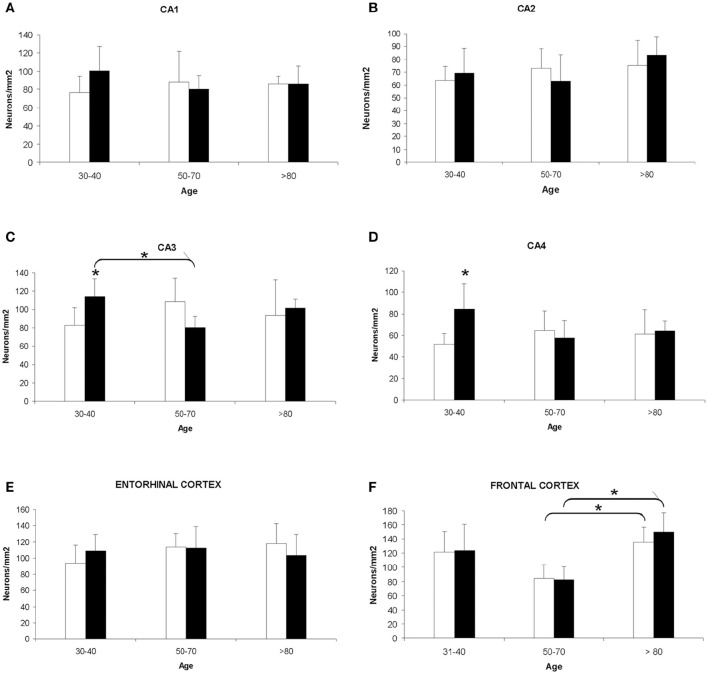
The aim of this study is the analysis of neuronal density in four areas of the hippocampus, and entorhinal and frontal cortices to analyze the possible gender influence during normal aging and in Alzheimer's disease (AD).
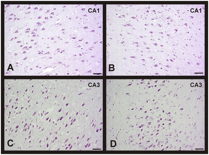
Human brain tissues of different age and from both sexes, without neurological pathology and with different Braak's stages of AD, were studied. Neuronal density was quantified using the optical dissector.
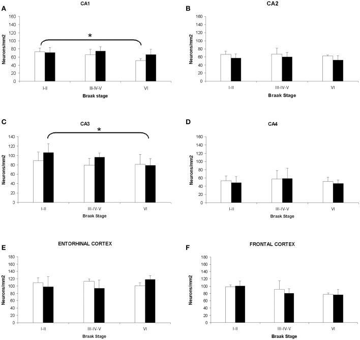
Our results showed the absence of a significant neuronal loss during aging in non-pathological brains in both sexes. However, we have demonstrated specific punctual significant variations in neuronal density related with the age and gender in some regions of these brains. In fact, we observed a higher neuronal density in CA3 and CA4 hippocampal areas of non-pathological brains of young men compared to women. During AD, we observed a negative correlation between Braak's stages and neuronal density in hippocampus, specifically in CA1 for women and CA3 for men, and in frontal cortex for both, men and women.
Our data demonstrated a sexual dimorphism in the neuronal vulnerability to degeneration suggesting the need to consider the gender of the individuals in future studies, regarding neuronal loss in aging and AD, in order to avoid problems in interpreting data.
学习过程或语言发展只是男性和女性在质上存在差异的部分认知功能。大脑结构的性别差异似乎是这些差异的背后原因。事实上,这种神经解剖学水平上的性二态性无疑伴随着衰老和神经退行性疾病对男性和女性大脑影响方式的差异。
本研究的目的是分析海马体四个区域、内嗅皮质和额叶皮质中的神经元密度,以分析正常衰老和阿尔茨海默病(AD)期间可能存在的性别影响。
研究了不同年龄、不同性别的无神经病理学病变且处于不同AD Braak阶段的人脑组织。使用光学分割器对神经元密度进行定量分析。
我们的结果显示,在非病理性大脑的衰老过程中,两性均未出现明显的神经元损失。然而,我们已经证明,在这些大脑的某些区域,神经元密度存在与年龄和性别相关的特定显著变化。事实上,我们观察到,与女性相比,年轻男性非病理性大脑的海马体CA3和CA4区域的神经元密度更高。在AD期间,我们观察到Braak阶段与海马体神经元密度之间存在负相关,具体而言,女性在CA1区域,男性在CA3区域,以及在男性和女性的额叶皮质中均如此。
我们的数据表明,在神经元对退化的易损性方面存在性二态性,这表明在未来关于衰老和AD中神经元损失的研究中,有必要考虑个体的性别,以避免在解释数据时出现问题。