Eyre Rachel, Alférez Denis G, Spence Kath, Kamal Mohamed, Shaw Frances L, Simões Bruno M, Santiago-Gómez Angélica, Sarmiento-Castro Aida, Bramley Maria, Absar Mohammed, Saad Zahida, Chatterjee Sumohan, Kirwan Cliona, Gandhi Ashu, Armstrong Anne C, Wardley Andrew M, O'Brien Ciara S, Farnie Gillian, Howell Sacha J, Clarke Robert B
Breast Biology Group, Breast Cancer Now Research Unit, Division of Molecular and Clinical Cancer Sciences, Manchester Cancer Research Centre, University of Manchester, Wilmslow Road, Manchester, M20 4QL, UK.
Department of Zoology, Faculty of Science, University of Benha, Benha, Egypt.
J Mammary Gland Biol Neoplasia. 2016 Dec;21(3-4):99-109. doi: 10.1007/s10911-016-9361-8. Epub 2016 Sep 28.
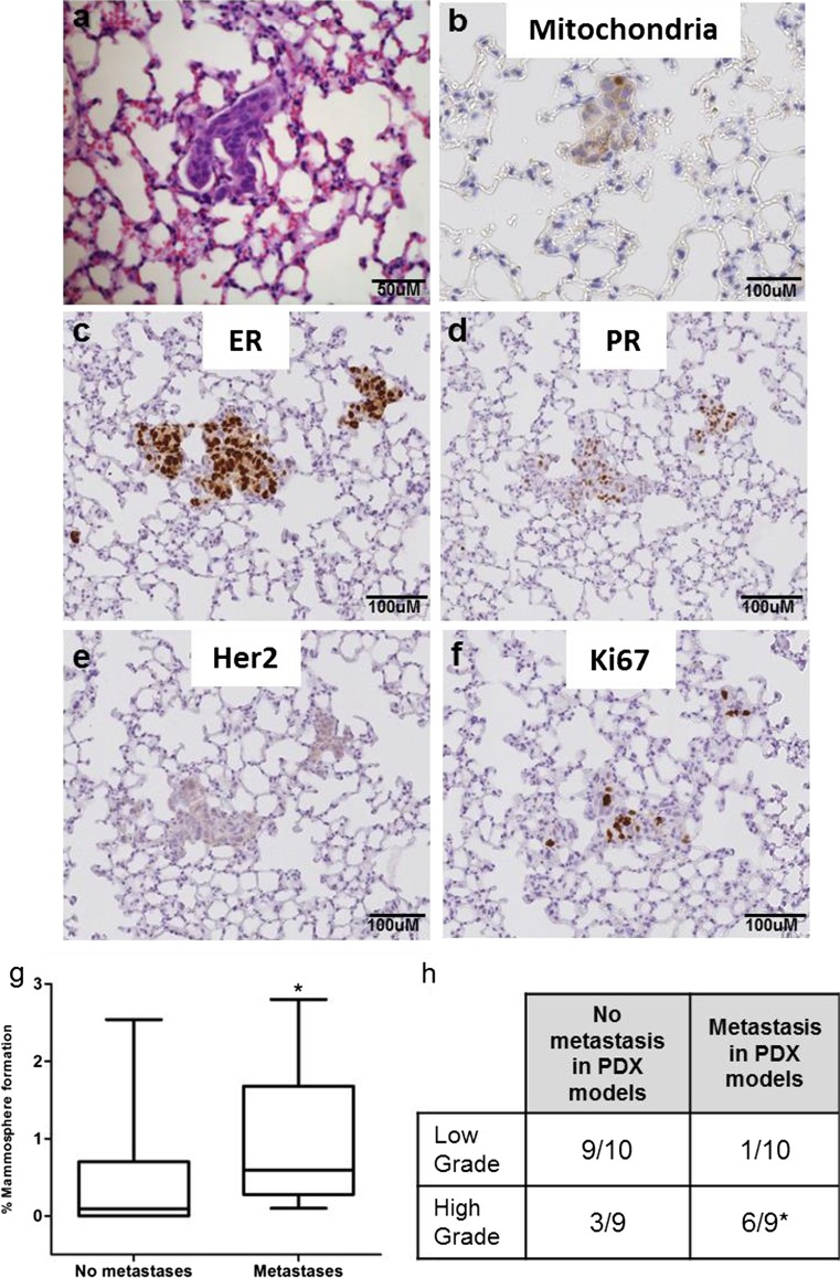
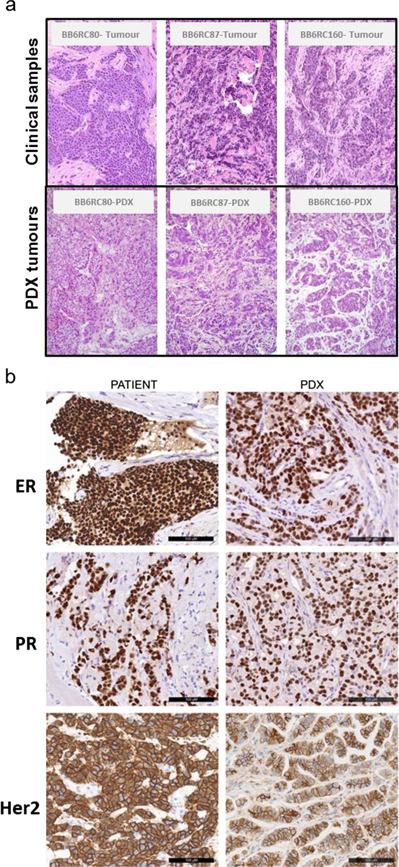
Breast cancer specific mortality results from tumour cell dissemination and metastatic colonisation. Identification of the cells and processes responsible for metastasis will enable better prevention and control of metastatic disease, thus reducing relapse and mortality. To better understand these processes, we prospectively collected 307 patient-derived breast cancer samples (n = 195 early breast cancers (EBC) and n = 112 metastatic samples (MBC)). We assessed colony-forming activity in vitro by growing isolated cells in both primary (formation) and secondary (self-renewal) mammosphere culture, and tumour initiating activity in vivo through subcutaneous transplantation of fragments or cells into mice. Metastatic samples formed primary mammosphere colonies significantly more frequently than early breast cancers and had significantly higher primary mammosphere colony formation efficiency (0.9 % vs. 0.6 %; p < 0.0001). Tumour initiation in vivo was significantly higher in metastatic than early breast cancer samples (63 % vs. 38 %, p = 0.04). Of 144 breast cancer samples implanted in vivo, we established 20 stable patient-derived xenograft (PDX) models at passage 2 or greater. Lung metastases were detected in mice from 14 PDX models. Mammosphere colony formation in vitro significantly correlated with the ability of a tumour to metastasise to the lungs in vivo (p = 0.05), but not with subcutaneous tumour initiation. In summary, the breast cancer stem cell activities of colony formation and tumour initiation are increased in metastatic compared to early samples, and predict metastasis in vivo. These results suggest that breast stem cell activity will predict for poor outcome tumours, and therapy targeting this activity will improve outcomes for patients with metastatic disease.
乳腺癌特异性死亡源于肿瘤细胞的播散和转移定植。识别负责转移的细胞和过程将有助于更好地预防和控制转移性疾病,从而降低复发率和死亡率。为了更好地理解这些过程,我们前瞻性地收集了307份患者来源的乳腺癌样本(n = 195例早期乳腺癌(EBC)和n = 112例转移样本(MBC))。我们通过在原发性(形成)和继发性(自我更新)乳腺球培养中培养分离的细胞来评估体外集落形成活性,并通过将片段或细胞皮下移植到小鼠体内来评估体内肿瘤起始活性。转移样本形成原发性乳腺球集落的频率明显高于早期乳腺癌,且原发性乳腺球集落形成效率显著更高(0.9%对0.6%;p < 0.0001)。转移性样本在体内的肿瘤起始能力明显高于早期乳腺癌样本(63%对38%,p = 0.04)。在144份植入体内的乳腺癌样本中,我们在传代2次或更高传代时建立了20个稳定的患者来源异种移植(PDX)模型。在14个PDX模型的小鼠中检测到肺转移。体外乳腺球集落形成与肿瘤在体内转移至肺的能力显著相关(p = 0.05),但与皮下肿瘤起始无关。总之,与早期样本相比,转移性样本中集落形成和肿瘤起始的乳腺癌干细胞活性增加,并可预测体内转移。这些结果表明,乳腺干细胞活性将预测预后不良的肿瘤,针对该活性的治疗将改善转移性疾病患者的预后。