Thakur Raghu Raj Singh, Tekko Ismaiel A, Al-Shammari Farhan, Ali Ahlam A, McCarthy Helen, Donnelly Ryan F
School of Pharmacy, Medical Biology Centre, Queen's University Belfast, 97 Lisburn Road, Belfast, BT9 7BL, Northern Ireland, UK.
Faculty of Pharmacy, Aleppo University, Aleppo, Syria.
Drug Deliv Transl Res. 2016 Dec;6(6):800-815. doi: 10.1007/s13346-016-0332-9.
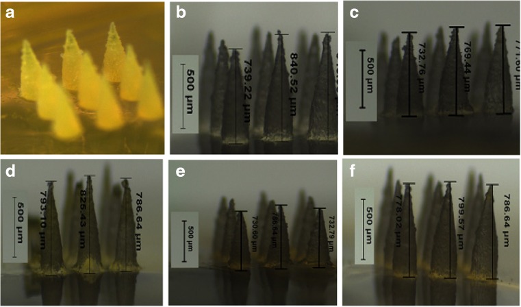
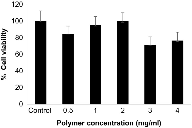
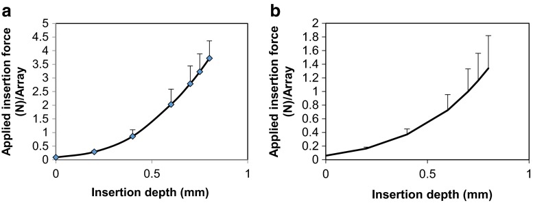
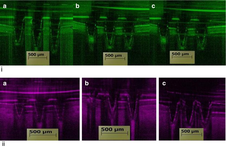
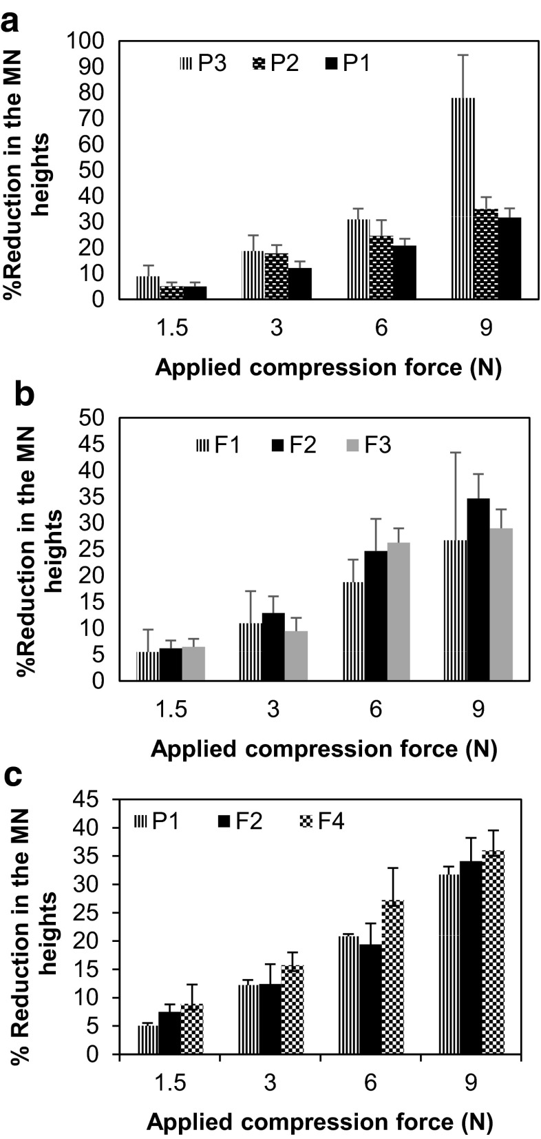
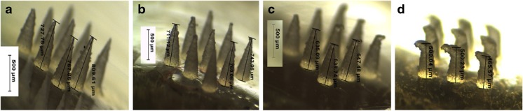
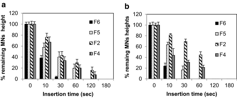
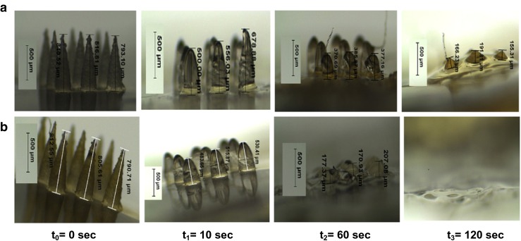
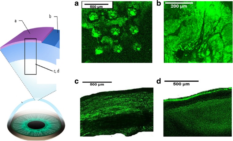
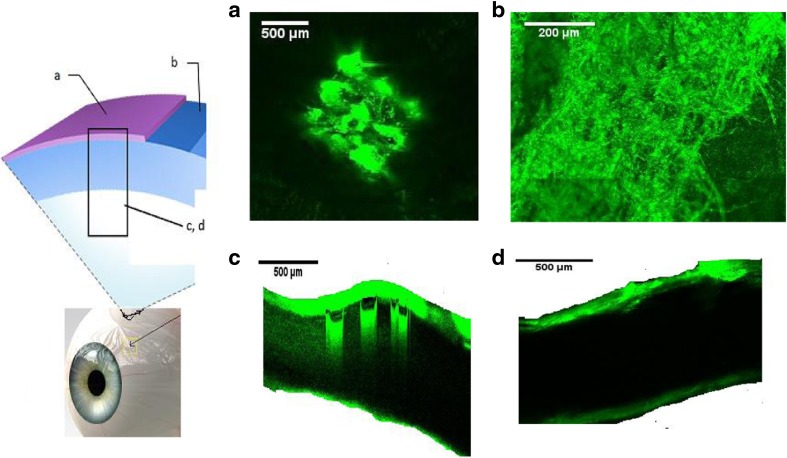
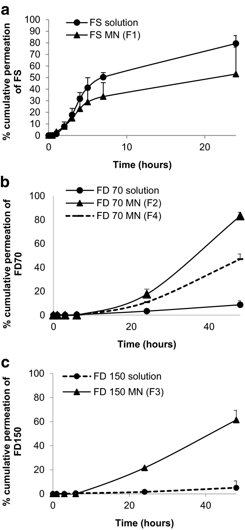
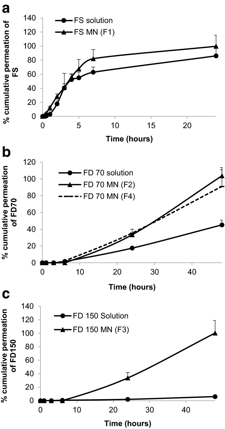
In this study, dissolving microneedles (MNs) were used to enhance ocular drug delivery of macromolecules. MNs were fabricated using polyvinylpyrrolidone (PVP) polymer of various molecular weights (MWs) containing three model molecules of increasing MW, namely fluorescein sodium and fluorescein isothiocyanate-dextrans (with MW of 70 k and 150 k Da). Arrays (3 × 3) of PVP MNs with conical shape measuring about 800 μm in height with a 300 μm base diameter, containing the model drugs, were fabricated and characterized for their fracture forces, insertion forces (in the sclera and cornea), depth of penetration (using OCT and confocal imaging), dissolution time and in vitro permeation. The average drug content of the MNs (only in MN shafts) ranged from 0.96 to 9.91 μg, and the average moisture content was below 11 %. High MW PVP produced MNs that can withstand higher forces with minimal reduction in needle height. PVP MNs showed rapid dissolution that ranged from 10 to 180 s, which was dependent upon PVP's MW. In vitro studies showed significant enhancement of macromolecule permeation when MNs were used, across both the corneal and scleral tissues, in comparison to topically applied aqueous solutions. Confocal images showed that the macromolecules formed depots within the tissues, which led to sustained permeation. However, use of MNs did not significantly benefit the permeation of small molecules; nevertheless, MN application has the potential for drug retention within the selected ocular tissues unlike topical application for small molecules. The material used in the fabrication of the MNs was found to be biocompatible with retinal cells (i.e. ARPE-19). Overall, this study reported the design and fabrication of minimally invasive rapidly dissolving polymeric MN arrays which were able to deliver high MW molecules to the eye via the intrastromal or intrascleral route. Thus, dissolving MNs have potential applications in enhancing ocular delivery of both small and macromolecules.
在本研究中,使用可溶解微针(MNs)来增强大分子药物的眼部递送。微针采用不同分子量(MW)的聚乙烯吡咯烷酮(PVP)聚合物制备,其中包含三种分子量递增的模型分子,即荧光素钠和异硫氰酸荧光素 - 葡聚糖(分子量分别为70k和150k Da)。制备了含有模型药物、高度约800μm、基部直径300μm的圆锥形PVP微针阵列(3×3),并对其断裂力、插入力(在巩膜和角膜中)、穿透深度(使用光学相干断层扫描(OCT)和共聚焦成像)、溶解时间和体外渗透进行了表征。微针(仅针杆部分)的平均药物含量在0.96至9.91μg之间,平均水分含量低于11%。高分子量PVP制备的微针能够承受更高的力,且针高降低最小。PVP微针的溶解速度很快,溶解时间在10至180秒之间,这取决于PVP的分子量。体外研究表明,与局部应用水溶液相比,使用微针时大分子在角膜和巩膜组织中的渗透均显著增强。共聚焦图像显示,大分子在组织内形成贮库,从而实现持续渗透。然而,微针的使用对小分子的渗透没有显著益处;尽管如此,与小分子的局部应用不同,微针应用有可能使药物保留在选定的眼部组织中。发现用于制造微针的材料与视网膜细胞(即ARPE - 19)具有生物相容性。总体而言,本研究报告了微创快速溶解聚合物微针阵列的设计与制造,该阵列能够通过基质内或巩膜内途径将高分子量分子递送至眼部。因此,可溶解微针在增强小分子和大分子的眼部递送方面具有潜在应用。