Yoshimitsu Kengo, Shinagawa Yoshinobu, Mitsufuji Toshimichi, Mutoh Emi, Urakawa Hiroshi, Sakamoto Keiko, Fujimitsu Ritsuko, Takano Koichi
Department of Radiology, Faculty of Medicine, Fukuoka University.
Magn Reson Med Sci. 2017 Jan 10;16(1):73-77. doi: 10.2463/mrms.mp.2016-0047. Epub 2016 Oct 11.
To elucidate whether any differences are present in the stiffness map obtained with a multiscale direct inversion algorithm (MSDI) vs that with a multimodel direct inversion algorithm (MMDI), both qualitatively and quantitatively.
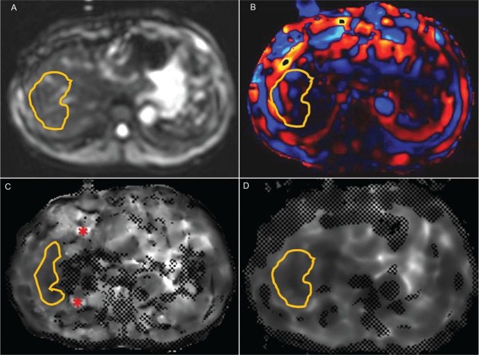
The MR elastography (MRE) data of 37 consecutive patients who underwent liver MR elastography between September and October 2014 were retrospectively analyzed by using both MSDI and MMDI. Two radiologists qualitatively assessed the stiffness maps for the image quality in consensus, and the measured liver stiffness and measurable areas were quantitatively compared between MSDI and MMDI.
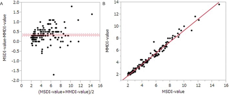
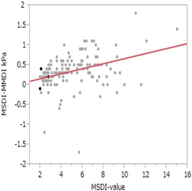
MMDI provided a stiffness map of better image quality, with comparable or slightly less artifacts. Measurable areas by MMDI (43.7 ± 17.8 cm) was larger than that by MSDI (37.5 ± 14.7 cm) (P < 0.05). Liver stiffness measured by MMDI (4.51 ± 2.32 kPa) was slightly (7%), but significantly less than that by MSDI (4.86 ± 2.44 kPa) (P < 0.05).
MMDI can provide stiffness map of better image quality, and slightly lower stiffness values as compared to MSDI at 3T MRE, which radiologists should be aware of.
从定性和定量两方面阐明采用多尺度直接反演算法(MSDI)获得的硬度图与采用多模型直接反演算法(MMDI)获得的硬度图是否存在差异。
回顾性分析2014年9月至10月间连续37例行肝脏磁共振弹性成像(MRE)患者的MRE数据,同时采用MSDI和MMDI进行分析。两名放射科医生对硬度图的图像质量进行了一致性定性评估,并对MSDI和MMDI之间测量的肝脏硬度和可测量区域进行了定量比较。
MMDI提供的硬度图图像质量更好,伪影相当或略少。MMDI的可测量区域(43.7±17.8 cm)大于MSDI的可测量区域(37.5±14.7 cm)(P<0.05)。MMDI测量的肝脏硬度(4.51±2.32 kPa)略低(7%),但显著低于MSDI测量的肝脏硬度(4.86±2.44 kPa)(P<0.05)。
在3T磁共振弹性成像中,MMDI能够提供图像质量更好的硬度图,且与MSDI相比,硬度值略低,放射科医生应予以注意。