Mizutani Ryuta, Saiga Rino, Ohtsuka Masato, Miura Hiromi, Hoshino Masato, Takeuchi Akihisa, Uesugi Kentaro
Department of Applied Biochemistry, Tokai University, Hiratsuka, Kanagawa 259-1292, Japan.
Tokai University School of Medicine, Isehara, Kanagawa 259-1193, Japan.
Sci Rep. 2016 Oct 11;6:35061. doi: 10.1038/srep35061.
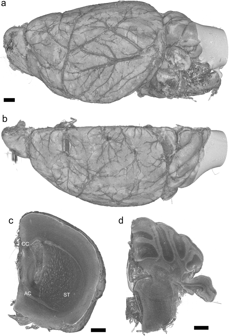
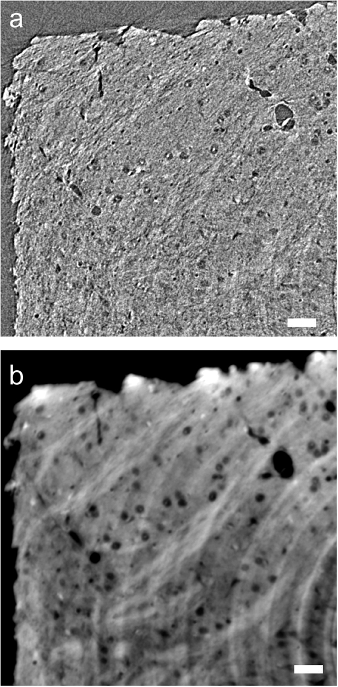
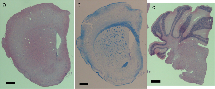
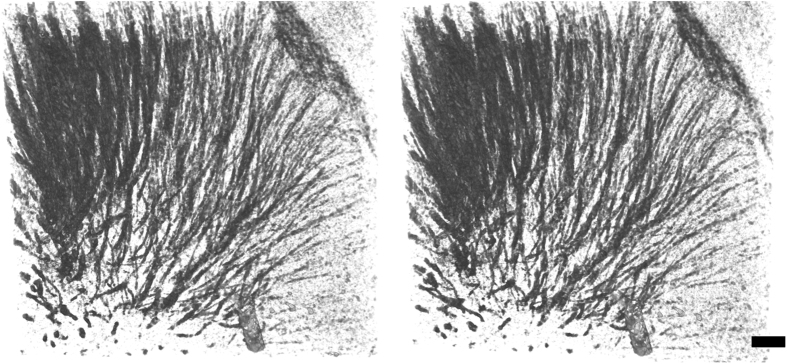
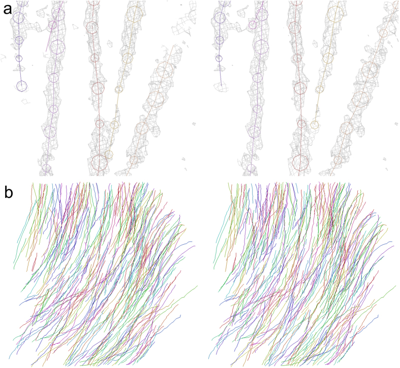
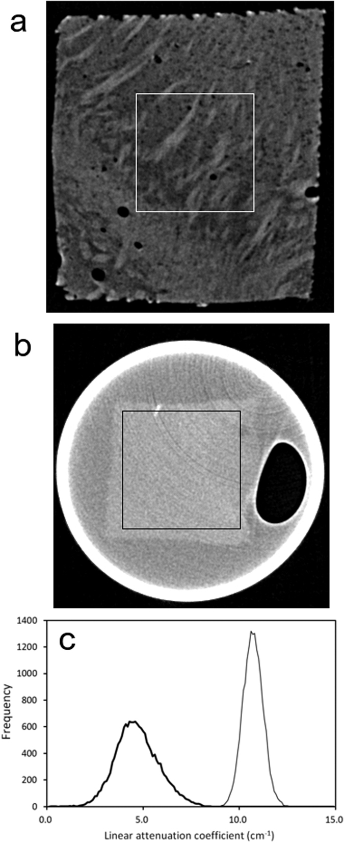
Neurons transmit active potentials through axons, which are essential for the brain to function. In this study, the axonal networks of the murine brain were visualized with X-ray tomographic microscopy, also known as X-ray microtomography or micro-CT. Murine brain samples were freeze-dried to reconstitute the intrinsic contrast of tissue constituents and subjected to X-ray visualization. A whole brain hemisphere visualized by absorption contrast illustrated three-dimensional structures including those of the striatum, corpus callosum, and anterior commissure. Axonal tracts observed in the striatum start from the basal surface of the cerebral cortex and end at various positions in the basal ganglia. The distribution of X-ray attenuation coefficients indicated that differences in water and phospholipid content between the myelin sheath and surrounding tissue constituents account for the observed contrast. A rod-shaped cutout of brain tissue was also analyzed with a phase retrieval method, wherein tissue microstructures could be resolved with up to 2.7 μm resolution. Structures of axonal networks of the striatum were reconstructed by tracing axonal tracts. Such an analysis should be able to delineate the functional relationships of the brain regions involved in the observed network.
神经元通过轴突传递动作电位,而轴突对于大脑发挥功能至关重要。在本研究中,利用X射线断层显微镜(也称为X射线显微断层成像或微CT)对小鼠大脑的轴突网络进行了可视化。将小鼠脑样本冷冻干燥以恢复组织成分的固有对比度,然后进行X射线可视化。通过吸收对比度可视化的整个脑半球展示了包括纹状体、胼胝体和前连合在内的三维结构。在纹状体中观察到的轴突束从大脑皮层的基底表面起始,终止于基底神经节的不同位置。X射线衰减系数的分布表明,髓鞘与周围组织成分之间水和磷脂含量的差异是观察到的对比度的原因。还采用相位恢复方法对脑组织的棒状切片进行了分析,其中组织微观结构的分辨率可达2.7μm。通过追踪轴突束重建了纹状体轴突网络的结构。这样的分析应该能够描绘出所观察到的网络中涉及的脑区之间的功能关系。