Liu Daihong, Duan Shanshan, Zhang Jiuquan, Zhou Chaoyang, Liang Minglong, Yin Xuntao, Wei Ping, Wang Jian
Department of Radiology, Southwest Hospital, Third Military Medical University Chongqing, China.
Department of Endocrinology, Southwest Hospital, Third Military Medical University Chongqing, China.
Front Hum Neurosci. 2016 Sep 27;10:490. doi: 10.3389/fnhum.2016.00490. eCollection 2016.
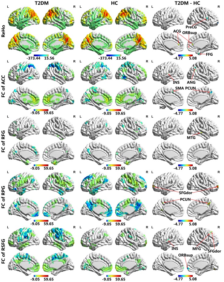
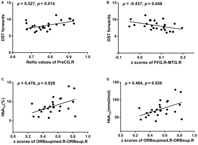
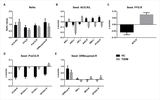
Type 2 diabetes mellitus (T2DM) has been associated with cognitive impairment. However, its neurological mechanism remains elusive. Combining regional homogeneity (ReHo) and functional connectivity (FC) analyses, the present study aimed to investigate brain functional alterations in middle-aged T2DM patients, which could provide complementary information for the neural substrates underlying T2DM-associated brain dysfunction. Twenty-five T2DM patients and 25 healthy controls were involved in neuropsychological testing and structural and resting-state functional magnetic resonance imaging (rs-fMRI) data acquisition. ReHo analysis was conducted to determine the peak coordinates of brain regions with abnormal local brain activity synchronization. Then, the identified brain regions were considered as seeds, and FC between these brain regions and global voxels was computed. Finally, the potential correlations between the imaging indices and neuropsychological data were also explored. Compared with healthy controls, T2DM patients exhibited higher ReHo values in the anterior cingulate gyrus (ACG) and lower ReHo in the right fusiform gyrus (FFG), right precentral gyrus (PreCG) and right medial orbit of the superior frontal gyrus (SFG). Considering these areas as seed regions, T2DM patients displayed aberrant FC, mainly in the frontal and parietal lobes. The pattern of FC alterations in T2DM patients was characterized by decreased connectivity and positive to negative or negative to positive converted connectivity. Digital Span Test (DST) forward scores revealed significant correlations with the ReHo values of the right PreCG (ρ = 0.527, = 0.014) and FC between the right FFG and middle temporal gyrus (MTG; ρ = -0.437, = 0.048). Our findings suggest that T2DM patients suffer from cognitive dysfunction related to spatially local and remote brain activity synchronization impairment. The patterns of ReHo and FC alterations shed light on the mechanisms underlying T2DM-associated brain dysfunction and might serve as imaging biomarkers for diagnosis and evaluation.
2型糖尿病(T2DM)与认知障碍有关。然而,其神经机制仍不清楚。本研究结合局部一致性(ReHo)和功能连接(FC)分析,旨在调查中年T2DM患者的脑功能改变,这可为T2DM相关脑功能障碍的神经基质提供补充信息。25例T2DM患者和25例健康对照者参与了神经心理学测试以及结构和静息态功能磁共振成像(rs-fMRI)数据采集。进行ReHo分析以确定局部脑活动同步异常的脑区的峰值坐标。然后,将识别出的脑区视为种子点,并计算这些脑区与全脑体素之间的FC。最后,还探讨了成像指标与神经心理学数据之间的潜在相关性。与健康对照相比,T2DM患者在前扣带回(ACG)的ReHo值较高,而在右侧梭状回(FFG)、右侧中央前回(PreCG)和右侧额上回内侧眶部(SFG)的ReHo值较低。将这些区域视为种子区,T2DM患者表现出异常的FC,主要在额叶和顶叶。T2DM患者的FC改变模式的特点是连接性降低以及连接性从正到负或从负到正的转换。数字广度测试(DST)正向得分与右侧PreCG的ReHo值(ρ = 0.527,P = 0.014)以及右侧FFG与颞中回(MTG)之间的FC(ρ = -0.437,P = 0.048)显著相关。我们的研究结果表明,T2DM患者存在与空间局部和远程脑活动同步受损相关的认知功能障碍。ReHo和FC改变模式揭示了T2DM相关脑功能障碍的潜在机制,并可能作为诊断和评估的影像学生物标志物。