Mielczarek Marzena, Michalska Joanna, Polatyńska Katarzyna, Olszewski Jurek
Department of Otolaryngology, Laryngological Oncology, Audiology, and Phoniatrics, Medical University of Lodz Lodz, Poland.
Department of Neurology, Polish Mother's Memorial Hospital Research Institute Lodz, Poland.
Front Neurosci. 2016 Oct 6;10:453. doi: 10.3389/fnins.2016.00453. eCollection 2016.
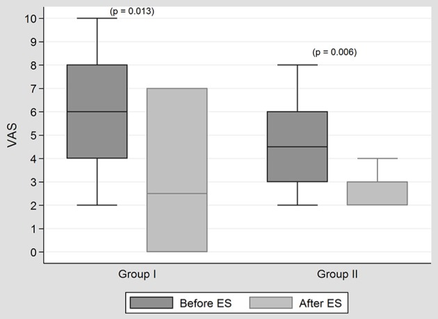
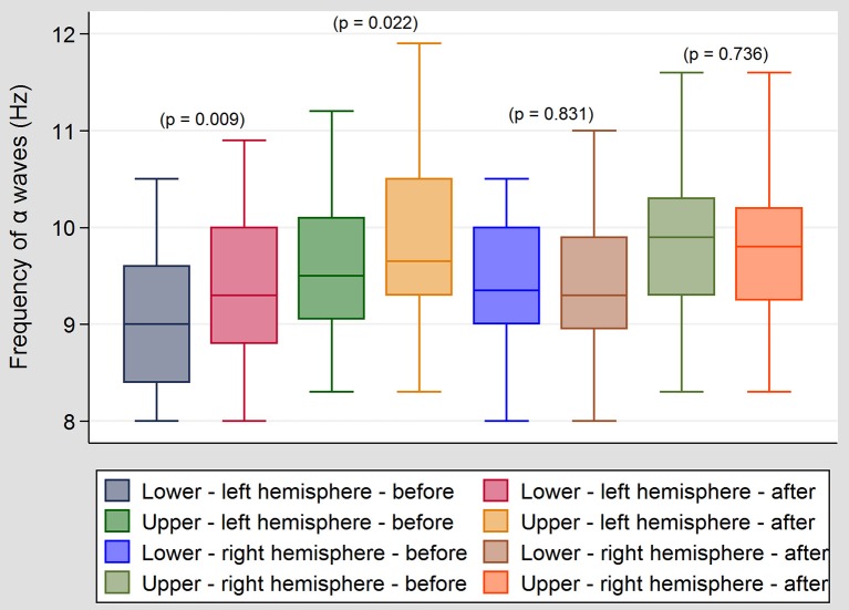
In our clinic invasive transtympanal promontory positive DC stimulations were first used, with a success rate of 42%. However, non-invasive hydrotransmissive negative DC stimulations are now favored, with improvement being obtained in 37.8% directly after the treatment, and 51.3% in a follow up 1 month after treatment. The further improvement after 1 month may be due to neuroplastic changes at central level as a result of altered peripheral input. The aim of the study was to determine how/whether a single electrical stimulation of the ear influences cortical activity, and whether changes observed in tinnitus after electrical stimulation are associated with any changes in cortical activity recorded in EEG. The study included 12 tinnitus patients (F-6, M-6) divided into two groups. Group I comprised six patients with unilateral tinnitus - unilateral, ipsilateral ES was performed. Group II comprised six patients with bilateral tinnitus-bilateral ES was performed. ES was performed using a custom-made apparatus. The active, silver probe-was immersed inside the external ear canal filled with saline. The passive electrode was placed on the forehead. The stimulating frequency was 250 Hz, the intensity ranged from 0.14 to 1.08 mA. The voltage was kept constant at 3 V. The duration of stimulation was 4 min. The EEG recording (Deymed QEST 32) was performed before and after ES. The patients assessed the intensity of tinnitus on the VAS 1-10. In both groups an improvement in VAS was observed-in group I-in five ears (83.3%), in group II-in seven ears (58.3%). In Group I, a significant increase in the upper and lower limit frequency of alpha band was observed in the central temporal and frontal regions following ES. These changes, however, were not correlated with improvement in tinnitus. No significant changes were observed in the beta and theta bands and in group II. Preliminary results of our research reveal a change in cortical activity after electrical stimulations of the ear. However, it remains unclear if it is primary or secondary to peripheral auditory excitation. No similar studies had been found in the literature.
在我们的诊所,首先使用了经鼓岬侵入性直流电刺激,成功率为42%。然而,现在更倾向于使用非侵入性水传导性负直流电刺激,治疗后直接改善的比例为37.8%,治疗后1个月随访时为51.3%。1个月后的进一步改善可能是由于外周输入改变导致中枢水平的神经可塑性变化。该研究的目的是确定单次耳部电刺激如何/是否影响皮层活动,以及电刺激后耳鸣的变化是否与脑电图记录的皮层活动变化相关。该研究包括12名耳鸣患者(女性6名,男性6名),分为两组。第一组包括6名单侧耳鸣患者——进行了单侧、同侧电刺激。第二组包括6名双侧耳鸣患者——进行了双侧电刺激。电刺激使用定制设备进行。活性银探头浸入充满盐水的外耳道内。无源电极置于前额。刺激频率为250Hz,强度范围为0.14至1.08mA。电压保持恒定在3V。刺激持续时间为4分钟。在电刺激前后进行脑电图记录(Deymed QEST 32)。患者在1-10视觉模拟量表上评估耳鸣强度。两组均观察到视觉模拟量表有所改善——第一组中5只耳朵(83.3%),第二组中7只耳朵(58.3%)。在第一组中,电刺激后中央颞区和额区的α波带上限和下限频率显著增加。然而,这些变化与耳鸣的改善无关。在第二组中,β波和θ波未观察到显著变化。我们研究的初步结果显示耳部电刺激后皮层活动发生了变化。然而,尚不清楚这是外周听觉兴奋的原发性还是继发性变化。文献中未发现类似研究。