Zheng Fang, Perlman Harris, Matthys Patrick, Wen Yurong, Lahoutte Tony, Muyldermans Serge, Lu Shemin, De Baetselier Patrick, Schoonooghe Steve, Devoogdt Nick, Raes Geert
Department of Biochemistry and Molecular Biology, Health Science Center, Xi'an Jiaotong University, Xi'an 710061, P. R. China.
Research group of Cellular and Molecular Immunology, Vrije Universiteit Brussel, Brussels, B-1050, Belgium.
Sci Rep. 2016 Oct 25;6:35966. doi: 10.1038/srep35966.
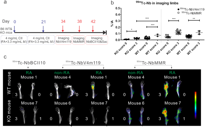
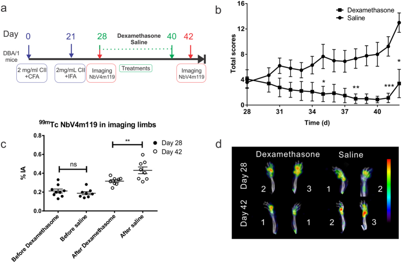
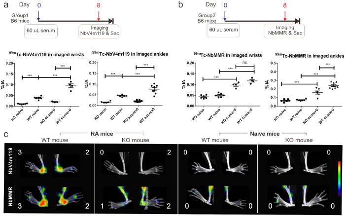
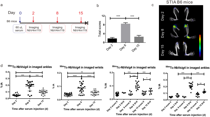
Single-photon emission computed tomography combined with micro-CT (SPECT/μCT) imaging using Nanobodies against complement receptor of the Ig superfamily (CRIg), found on tissue macrophages such as synovial macrophages, has promising potential to visualize joint inflammation in experimental arthritis. Here, we further addressed the specificity and assessed the potential for arthritis monitoring. Signals obtained with Tc-labelled NbV4m119 Nanobody were compared in joints of wild type (WT) versus CRIg mice with collagen-induced arthritis (CIA) or K/BxN serum transfer-induced arthritis (STIA). In addition, SPECT/μCT imaging was used to investigate arthritis development in STIA and in CIA under dexamethasone treatment. Tc-NbV4m119 accumulated in inflamed joints of WT, but not CRIg mice with CIA and STIA. Development and spontaneous recovery of symptoms in STIA was reflected in initially increased and subsequently reduced joint accumulation of Tc-NbV4m119. Dexamethasone treatment of CIA mice reduced Tc-NbV4m119 accumulation as compared to saline control in most joints except knees. SPECT/μCT imaging with Tc-NbV4m119 allows specific assessment of inflammation in different arthritis models and provides complementary information to clinical scoring for quantitatively and non-invasively monitoring the pathological process and the efficacy of arthritis treatment.
使用针对免疫球蛋白超家族补体受体(CRIg)的纳米抗体进行单光子发射计算机断层扫描与微型计算机断层扫描(SPECT/μCT)联合成像,CRIg存在于诸如滑膜巨噬细胞等组织巨噬细胞上,在可视化实验性关节炎中的关节炎症方面具有广阔的潜力。在此,我们进一步探讨了其特异性,并评估了其用于关节炎监测的潜力。将用锝标记的NbV4m119纳米抗体获得的信号在野生型(WT)小鼠与患有胶原诱导性关节炎(CIA)或K/BxN血清转移诱导性关节炎(STIA)的CRIg小鼠的关节中进行比较。此外,利用SPECT/μCT成像研究了地塞米松治疗下STIA和CIA的关节炎发展情况。Tc-NbV4m119在患有CIA和STIA的WT小鼠的炎症关节中积聚,但在CRIg小鼠中不积聚。STIA症状的发展和自发恢复反映在Tc-NbV4m119在关节中的积聚最初增加,随后减少。与生理盐水对照相比,地塞米松治疗CIA小鼠可减少除膝盖外大多数关节中Tc-NbV4m119的积聚。用Tc-NbV4m119进行SPECT/μCT成像能够特异性评估不同关节炎模型中的炎症,并为临床评分提供补充信息,用于定量和非侵入性监测病理过程及关节炎治疗效果。