Ueno Takuya, Kim Pilhan, McGrath Martina M, Yeung Melissa Y, Shimizu Tetsunosuke, Jung Keehoon, Sayegh Mohamed H, Chandraker Anil K, Abdi Reza, Yun Seok H
Renal Division, Transplantation Research Center, Brigham and Women's Hospital, Harvard Medical School , Boston, MA , USA.
Department of Dermatology, Wellman Center for Photomedicine, Massachusetts General Hospital, Harvard Medical School , Boston, MA , USA.
Front Immunol. 2016 Oct 14;7:412. doi: 10.3389/fimmu.2016.00412. eCollection 2016.
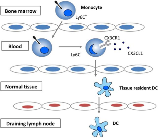
A number of studies have demonstrated the role of CX3CR1 in regulating the migration of monocytes into peripheral tissue and their transformation into dendritic cell (DC). No data are yet available on the importance of chemokine pathways in regulating homeostasis of DC in heart transplants. Recently, we showed that recipients of heart allografts from CX3CR1 donors show longer survival. To assess the trafficking of dDC, we have developed and tested a novel imaging tool in CX3CR1 DC (B6 background) heart graft into BALB/c recipient model.
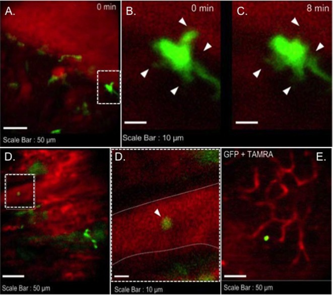
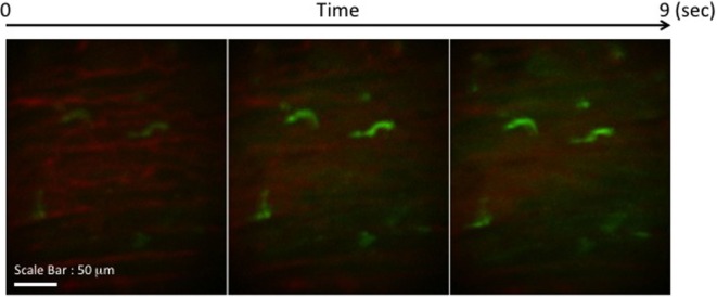
Majority of GFP cells were noted in the middle of cardiac myocyte. However few hours post transplant, they experienced morphological changes including stretching their extensions (3 and 24 h). However, images from 72 h at cardiac graft showed many of GFP cells moved to vessel areas. GFP cells were detected in near vessel wall. Only one GFP cell was observed in three lymph nodes (two mesenteric and one inguinal) (72 h).
Our data indicate that immediately post transplant dDC undergo morphological changes and traffic out of the organs via systemic circulation. While, we still noted presence of dDC in the transplanted organs, their trafficking to lymphoid tissue remains to be fully explored.
多项研究已证明CX3CR1在调节单核细胞向外周组织迁移及其向树突状细胞(DC)转化中的作用。关于趋化因子途径在调节心脏移植中DC稳态的重要性,目前尚无相关数据。最近,我们发现来自CX3CR1供体的心脏同种异体移植受者存活时间更长。为了评估树突状DC的迁移情况,我们开发并测试了一种新型成像工具,用于观察CX3CR1 DC(B6背景)心脏移植到BALB/c受体模型中的情况。
大多数绿色荧光蛋白(GFP)细胞出现在心肌细胞中部。然而,移植后数小时,它们经历了形态变化,包括伸展其突起(3小时和24小时)。然而,心脏移植72小时后的图像显示,许多GFP细胞迁移到血管区域。在血管壁附近检测到GFP细胞。在三个淋巴结(两个肠系膜淋巴结和一个腹股沟淋巴结)中仅观察到一个GFP细胞(72小时)。
我们的数据表明,移植后即刻,树突状DC会发生形态变化,并通过体循环离开器官。虽然我们仍注意到移植器官中存在树突状DC,但其向淋巴组织的迁移情况仍有待充分探索。