• 文献检索
  • 文档翻译
  • 深度研究
  • 学术资讯
  • Suppr Zotero 插件Zotero 插件
  • 邀请有礼
  • 套餐&价格
  • 历史记录
应用&插件
Suppr Zotero 插件Zotero 插件浏览器插件Mac 客户端Windows 客户端微信小程序
定价
高级版会员购买积分包购买API积分包
服务
文献检索文档翻译深度研究API 文档MCP 服务
关于我们
关于 Suppr公司介绍联系我们用户协议隐私条款
关注我们

Suppr 超能文献

核心技术专利:CN118964589B侵权必究
粤ICP备2023148730 号-1Suppr @ 2026

文献检索

告别复杂PubMed语法,用中文像聊天一样搜索,搜遍4000万医学文献。AI智能推荐,让科研检索更轻松。

立即免费搜索

文件翻译

保留排版,准确专业,支持PDF/Word/PPT等文件格式,支持 12+语言互译。

免费翻译文档

深度研究

AI帮你快速写综述,25分钟生成高质量综述,智能提取关键信息,辅助科研写作。

立即免费体验

从右心室等容收缩开始至S波峰值的时间:一种用于无创评估肺动脉高压的新组织多普勒成像指标。

Time From the Beginning of the Right Ventricle Isovolumetric Contraction to the Peak of the S Wave: A New TDI Indicator for the Non-Invasive Estimation of Pulmonary Hypertension.

作者信息

Parsaee Mozhgan, Ghaderi Fereshteh, Alizadehasl Azin, Bakhshandeh Hooman

机构信息

Echocardiography Research Center, Rajaie Cardiovascular Medical and Research Center, Iran University of Medical Sciences, Tehran, IR Iran.

Department of Cardiovascular Medicine, Echocardiography laboratory, Atherosclerosis Prevention Research Center, School of Medicine, Mashhad University of Medical Sciences, Mashhad, IR Iran.

出版信息

Res Cardiovasc Med. 2016 Jul 20;5(3):e26494. doi: 10.5812/cardiovascmed.26494. eCollection 2016 Aug.

DOI:10.5812/cardiovascmed.26494
PMID:27800451
原文链接:https://pmc.ncbi.nlm.nih.gov/articles/PMC5075392/
Abstract

BACKGROUND

Echocardiography is a key screening tool in the diagnostic algorithm of pulmonary hypertension (PH). In addition, tissue doppler imaging (TDI) is a promising method for the noninvasive estimation of pulmonary artery pressure (PAP).

OBJECTIVES

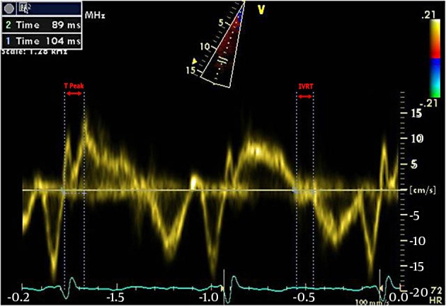
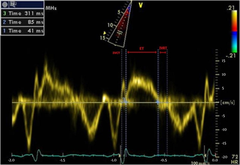
The aim of this study was to validate the accuracy of measuring the time from the beginning of the right ventricular isovolumetric contraction time (RV IVRT) to the peak of the S wave in the TDI of the base of the RV free wall (time to peak or TTP), as an indicator for the non-invasive estimation of pulmonary hypertension.

PATIENTS AND METHODS

In this diagnostic test study, 60 consecutive patients referred for right heart catheterization (RHC) were enrolled. A pulse-wave TDI was performed before the cardiac catheterization, with a mean interval of 1 hour between the two measurements. The TDI variables, such as the RV IVRT, myocardial performance index (MPI), and the new "time to peak" parameter, were measured at the lateral basal RV free wall. The patients were divided into two sub-groups according to the RHC findings: no-PH (mean PAP < 25 mmHg) and PH (mean PAP ≥ 25 mmHg) groups. Then, we calculated the specificity and sensitivity of the TDI parameters (including the TTP) for the diagnosis of PH.

RESULTS

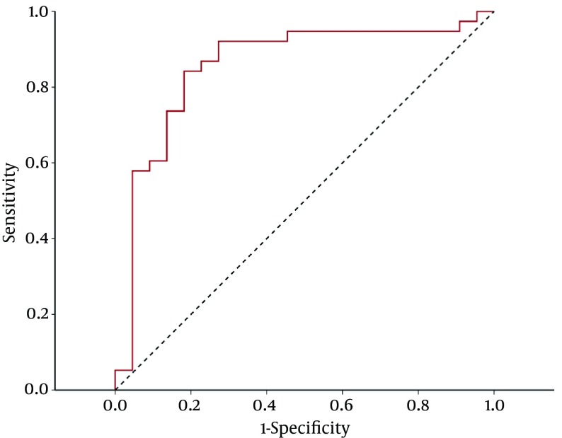
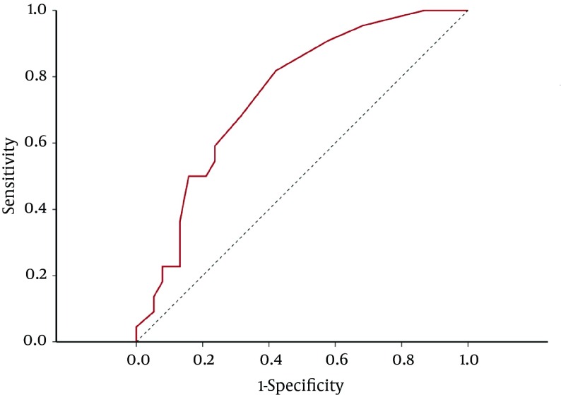
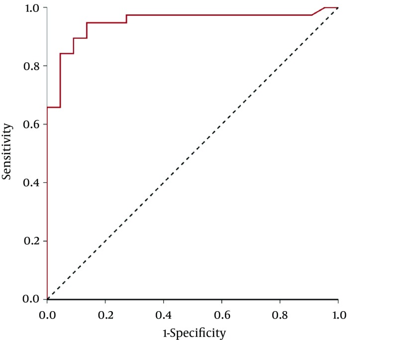
In our study, the TTP showed a significant inverse relationship with the PAP. Based on our results, a TTP of less than 127 ms could be used to predict PH, with a sensitivity and specificity of about 70% (AUC = 0.746 ± 0.064).

CONCLUSIONS

Based on the results of this study, we suggest the use of a novel "time from the beginning of isovolumetric contraction to the peak of the S wave" (TTP) parameter in the TDI of the base of the RV free wall to predict PH with acceptable accuracy in comparison with RHC.

摘要

背景

超声心动图是肺动脉高压(PH)诊断流程中的关键筛查工具。此外,组织多普勒成像(TDI)是一种用于无创估计肺动脉压(PAP)的有前景的方法。

目的

本研究的目的是验证测量从右心室等容收缩时间(RV IVRT)开始至右心室游离壁基部TDI中S波峰值的时间(达到峰值时间或TTP)作为无创估计肺动脉高压指标的准确性。

患者与方法

在这项诊断性试验研究中,纳入了60例连续接受右心导管检查(RHC)的患者。在心脏导管检查前进行脉冲波TDI,两次测量之间的平均间隔为1小时。在右心室游离壁外侧基部测量TDI变量,如RV IVRT、心肌性能指数(MPI)和新的“达到峰值时间”参数。根据RHC结果将患者分为两个亚组:无PH(平均PAP < 25 mmHg)和PH(平均PAP≥25 mmHg)组。然后,我们计算了TDI参数(包括TTP)对PH诊断的特异性和敏感性。

结果

在我们的研究中,TTP与PAP呈显著负相关。根据我们的结果,TTP小于127 ms可用于预测PH,敏感性和特异性约为70%(AUC = 0.746±0.064)。

结论

基于本研究结果,我们建议在右心室游离壁基部的TDI中使用一种新的“从等容收缩开始至S波峰值的时间”(TTP)参数,与RHC相比,以可接受的准确性预测PH。

https://cdn.ncbi.nlm.nih.gov/pmc/blobs/2e3c/5075392/e3c071b0b4cc/cardiovascmed-05-03-26494-i003.jpg
https://cdn.ncbi.nlm.nih.gov/pmc/blobs/2e3c/5075392/38894bd3f88c/cardiovascmed-05-03-26494-g001.jpg
https://cdn.ncbi.nlm.nih.gov/pmc/blobs/2e3c/5075392/4e3712d79099/cardiovascmed-05-03-26494-g002.jpg
https://cdn.ncbi.nlm.nih.gov/pmc/blobs/2e3c/5075392/9f958bb0d4d2/cardiovascmed-05-03-26494-i001.jpg
https://cdn.ncbi.nlm.nih.gov/pmc/blobs/2e3c/5075392/c27950a1ada7/cardiovascmed-05-03-26494-i002.jpg
https://cdn.ncbi.nlm.nih.gov/pmc/blobs/2e3c/5075392/e3c071b0b4cc/cardiovascmed-05-03-26494-i003.jpg

相似文献

1
Time From the Beginning of the Right Ventricle Isovolumetric Contraction to the Peak of the S Wave: A New TDI Indicator for the Non-Invasive Estimation of Pulmonary Hypertension.从右心室等容收缩开始至S波峰值的时间:一种用于无创评估肺动脉高压的新组织多普勒成像指标。
Res Cardiovasc Med. 2016 Jul 20;5(3):e26494. doi: 10.5812/cardiovascmed.26494. eCollection 2016 Aug.
2
Right Ventricular Tissue Doppler Myocardial Performance Index in Children with Pulmonary Hypertension: Relation to Invasive Hemodynamics.肺动脉高压患儿右心室组织多普勒心肌做功指数与有创血流动力学的关系
Pediatr Cardiol. 2018 Jan;39(1):98-104. doi: 10.1007/s00246-017-1733-3. Epub 2017 Oct 4.
3
Does the myocardial performance index affect pulmonary artery pressure in patients with mitral stenosis? A tissue Doppler imaging study.心肌性能指数是否影响二尖瓣狭窄患者的肺动脉压?一项组织多普勒成像研究。
Echocardiography. 2003 Apr;20(3):249-56. doi: 10.1046/j.1540-8175.2003.03022.x.
4
Noninvasive model including right ventricular speckle tracking for the evaluation of pulmonary hypertension.用于评估肺动脉高压的无创模型,包括右心室斑点追踪技术。
World J Cardiol. 2016 Aug 26;8(8):472-80. doi: 10.4330/wjc.v8.i8.472.
5
RV contractility and exercise-induced pulmonary hypertension in chronic mountain sickness: a stress echocardiographic and tissue Doppler imaging study.慢性高原病患者右心室收缩功能与运动诱发肺动脉高压:一项超声心动图及组织多普勒成像研究。
JACC Cardiovasc Imaging. 2013 Dec;6(12):1287-97. doi: 10.1016/j.jcmg.2013.08.007. Epub 2013 Oct 23.
6
Usefulness of tissue Doppler imaging to predict arrhythmic events in adults with repaired tetralogy of Fallot.组织多普勒成像预测法洛四联症修复术后成年人心律失常事件的效用。
Rev Port Cardiol. 2010 Jul-Aug;29(7-8):1145-61.
7
Right ventricular dysfunction and remodeling in chronic obstructive pulmonary disease without pulmonary hypertension.慢性阻塞性肺疾病患者中无肺动脉高压时的右心室功能障碍和重构。
J Am Coll Cardiol. 2013 Sep 17;62(12):1103-1111. doi: 10.1016/j.jacc.2013.04.091. Epub 2013 Jul 10.
8
Right ventricle performances with echocardiography and Tc myocardial perfusion imaging in pulmonary arterial hypertension patients.超声心动图与 Tc 心肌灌注显像评估肺动脉高压患者右心室功能。
Exp Biol Med (Maywood). 2018 May;243(9):754-761. doi: 10.1177/1535370218775321.
9
The impact of hypertension and hypertension-related left ventricle hypertrophy on right ventricle function.高血压及高血压相关左心室肥厚对右心室功能的影响。
Echocardiography. 2007 Apr;24(4):374-84. doi: 10.1111/j.1540-8175.2007.00419.x.
10
Pulmonary hypertension and left heart function: insights from tissue Doppler imaging and myocardial performance index.肺动脉高压与左心功能:组织多普勒成像和心肌性能指数的见解
Echocardiography. 2007 Apr;24(4):366-73. doi: 10.1111/j.1540-8175.2007.00405.x.

引用本文的文献

1
Right heart echocardiography findings in hypoxic pneumonia patients during the COVID-19 pandemic in a South African population.南非人群中新冠疫情期间缺氧性肺炎患者的右心超声心动图检查结果
Eur Heart J Imaging Methods Pract. 2023 Oct 25;1(2):qyad030. doi: 10.1093/ehjimp/qyad030. eCollection 2023 Sep.
2
Isovolumetric Contraction as a Marker of Ventricular Performance in Patients with Afterload Mismatch.等容收缩作为后负荷不匹配患者心室功能的标志物
Diagnostics (Basel). 2023 Apr 7;13(8):1366. doi: 10.3390/diagnostics13081366.
3
Doppler trans-thoracic echocardiography for detection of pulmonary hypertension in adults.

本文引用的文献

1
Definitions and diagnosis of pulmonary hypertension.肺动脉高压的定义和诊断。
J Am Coll Cardiol. 2013 Dec 24;62(25 Suppl):D42-50. doi: 10.1016/j.jacc.2013.10.032.
2
Right ventricular dysfunction and remodeling in chronic obstructive pulmonary disease without pulmonary hypertension.慢性阻塞性肺疾病患者中无肺动脉高压时的右心室功能障碍和重构。
J Am Coll Cardiol. 2013 Sep 17;62(12):1103-1111. doi: 10.1016/j.jacc.2013.04.091. Epub 2013 Jul 10.
3
Can pulmonary hypertension and increased pulmonary vascular resistance be ruled in and ruled out by echocardiography?
经胸多普勒超声心动图用于检测成人肺动脉高压。
Cochrane Database Syst Rev. 2022 May 9;5(5):CD012809. doi: 10.1002/14651858.CD012809.pub2.
超声心动图能否诊断和排除肺动脉高压和肺血管阻力增加?
J Am Soc Echocardiogr. 2013 May;26(5):469-78. doi: 10.1016/j.echo.2013.02.011. Epub 2013 Mar 16.
4
Derivation of mean pulmonary artery pressure from noninvasive parameters.从无创参数推导平均肺动脉压。
J Am Soc Echocardiogr. 2013 May;26(5):464-8. doi: 10.1016/j.echo.2013.01.006. Epub 2013 Feb 11.
5
Right isovolumic contraction velocity predicts survival in pulmonary hypertension.右室等容收缩速度可预测肺动脉高压患者的生存情况。
J Am Soc Echocardiogr. 2013 Mar;26(3):297-306. doi: 10.1016/j.echo.2012.11.011. Epub 2012 Dec 21.
6
Echocardiography in pulmonary arterial hypertension: from diagnosis to prognosis.超声心动图在肺动脉高压中的应用:从诊断到预后。
J Am Soc Echocardiogr. 2013 Jan;26(1):1-14. doi: 10.1016/j.echo.2012.10.009. Epub 2012 Nov 8.
7
Echocardiographic assessment of pulmonary hypertension: standard operating procedure.超声心动图评估肺动脉高压:标准操作流程。
Eur Respir Rev. 2012 Sep 1;21(125):239-48. doi: 10.1183/09059180.00003912.
8
Validation of the isovolumetric relaxation time for the estimation of pulmonary systolic arterial blood pressure in chronic pulmonary hypertension.等容舒张时间对慢性肺动脉高压患者肺动脉收缩压估测的验证。
Eur Heart J Cardiovasc Imaging. 2013 Jan;14(1):51-5. doi: 10.1093/ehjci/jes093. Epub 2012 May 15.
9
Assessment of right ventricular structure and function in pulmonary hypertension.肺动脉高压时右心室结构和功能的评估
J Cardiovasc Ultrasound. 2011 Sep;19(3):115-25. doi: 10.4250/jcu.2011.19.3.115. Epub 2011 Sep 30.
10
Echocardiographic indexes for the non-invasive evaluation of pulmonary hemodynamics.超声心动图指标用于肺循环血流动力学的无创评估。
J Am Soc Echocardiogr. 2010 Mar;23(3):225-39; quiz 332-4. doi: 10.1016/j.echo.2010.01.003.