• 文献检索
  • 文档翻译
  • 深度研究
  • 学术资讯
  • Suppr Zotero 插件Zotero 插件
  • 邀请有礼
  • 套餐&价格
  • 历史记录
应用&插件
Suppr Zotero 插件Zotero 插件浏览器插件Mac 客户端Windows 客户端微信小程序
定价
高级版会员购买积分包购买API积分包
服务
文献检索文档翻译深度研究API 文档MCP 服务
关于我们
关于 Suppr公司介绍联系我们用户协议隐私条款
关注我们

Suppr 超能文献

核心技术专利:CN118964589B侵权必究
粤ICP备2023148730 号-1Suppr @ 2026

文献检索

告别复杂PubMed语法,用中文像聊天一样搜索,搜遍4000万医学文献。AI智能推荐,让科研检索更轻松。

立即免费搜索

文件翻译

保留排版,准确专业,支持PDF/Word/PPT等文件格式,支持 12+语言互译。

免费翻译文档

深度研究

AI帮你快速写综述,25分钟生成高质量综述,智能提取关键信息,辅助科研写作。

立即免费体验

无标记成像技术在活细胞中检测组氨酸介导的 G 蛋白偶联受体激活。

Label-Free Imaging of Histamine Mediated G Protein-Coupled Receptors Activation in Live Cells.

机构信息

Biodesign Center for Bioelectronics and Biosensors, Arizona State University , Tempe, Arizona 85287, United States.

Department of Chemistry, Beijing Key Laboratory for Microanalytical Methods and Instrumentation, Tsinghua University , Beijing 100084, China.

出版信息

Anal Chem. 2016 Dec 6;88(23):11498-11503. doi: 10.1021/acs.analchem.6b02677. Epub 2016 Nov 15.

DOI:10.1021/acs.analchem.6b02677
PMID:27802015
原文链接:https://pmc.ncbi.nlm.nih.gov/articles/PMC5144837/
Abstract

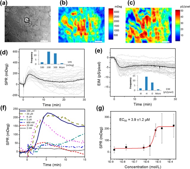
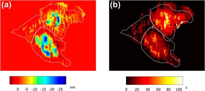
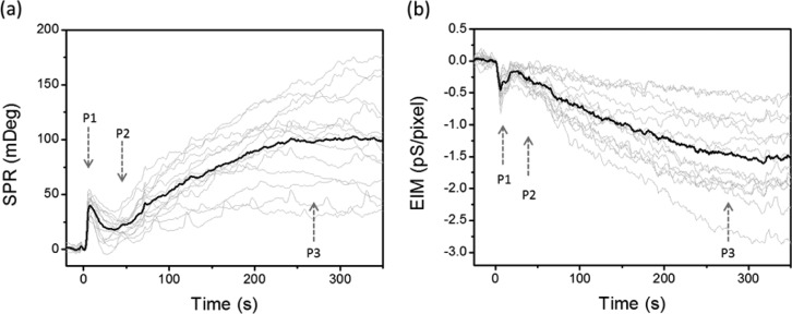
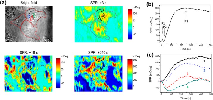
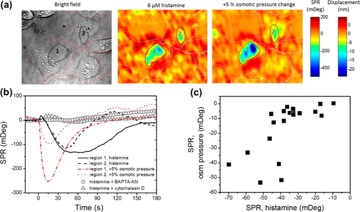
G protein-coupled receptors (GPCRs) are the largest protein family for cell signal transduction, and most of them are crucial drug targets. Conventional label-free assays lack the spatial information to address the heterogeneous response from single cells after GPCRs activation. Here, we reported a GPCRs study in live cells using plasmonic-based electrochemical impedance microscopy. This label-free optical imaging platform is able to resolve responses from individual cells with subcellular resolution. Using this platform, we studied the histamine mediated GPCRs activation and revealed spatiotemporal heterogeneity of cellular downstream responses. Triphasic responses were observed from individual HeLa cells upon histamine stimulation. A quick peak P1 in less than 10 s was attributed to the GPCRs triggered calcium release. An inverted P2 phase within 1 min was attributed to the alternations of cell-matrix adhesion after the activation of Protein Kinase C (PKC). The main peak (P3) around 3-6 min after the histamine treatment was due to dynamic mass redistribution and showed a dose-dependent response with a half-maximal effective concentration (EC) of 3.9 ± 1.2 μM. Heterogeneous P3 responses among individual cells were observed, particularly at high histamine concentration, indicating diverse histamine H1 receptor expression level in the cell population.

摘要

G 蛋白偶联受体(GPCRs)是细胞信号转导中最大的蛋白质家族,其中大多数是至关重要的药物靶点。传统的无标记检测方法缺乏空间信息,无法解决 GPCR 激活后单个细胞的异质反应。在这里,我们使用基于等离子体的电化学阻抗显微镜报告了活细胞中的 GPCRs 研究。这个无标记的光学成像平台能够以亚细胞分辨率解析来自单个细胞的反应。使用该平台,我们研究了组胺介导的 GPCRs 激活,并揭示了细胞下游反应的时空异质性。组胺刺激单个 HeLa 细胞时观察到三相反应。不到 10 秒的快速峰 P1 归因于 GPCR 触发的钙释放。1 分钟内的倒置 P2 相归因于蛋白激酶 C(PKC)激活后细胞-基质粘附的改变。组胺处理后 3-6 分钟左右的主要峰(P3)是由于动态质量重新分布,并且表现出与半最大有效浓度(EC)为 3.9 ± 1.2 μM 的剂量依赖性反应。在高组胺浓度下观察到单个细胞之间的异质 P3 反应,表明细胞群体中存在不同的组胺 H1 受体表达水平。

https://cdn.ncbi.nlm.nih.gov/pmc/blobs/8927/5144837/85304117357d/ac-2016-02677h_0004.jpg
https://cdn.ncbi.nlm.nih.gov/pmc/blobs/8927/5144837/babdd12c4081/ac-2016-02677h_0007.jpg
https://cdn.ncbi.nlm.nih.gov/pmc/blobs/8927/5144837/1c157ae701ad/ac-2016-02677h_0001.jpg
https://cdn.ncbi.nlm.nih.gov/pmc/blobs/8927/5144837/60e0865306a0/ac-2016-02677h_0002.jpg
https://cdn.ncbi.nlm.nih.gov/pmc/blobs/8927/5144837/26d134ef73d3/ac-2016-02677h_0003.jpg
https://cdn.ncbi.nlm.nih.gov/pmc/blobs/8927/5144837/85304117357d/ac-2016-02677h_0004.jpg

相似文献

1
Label-Free Imaging of Histamine Mediated G Protein-Coupled Receptors Activation in Live Cells.无标记成像技术在活细胞中检测组氨酸介导的 G 蛋白偶联受体激活。
Anal Chem. 2016 Dec 6;88(23):11498-11503. doi: 10.1021/acs.analchem.6b02677. Epub 2016 Nov 15.
2
Real-time monitoring of morphological changes in living cells by electronic cell sensor arrays: an approach to study G protein-coupled receptors.利用电子细胞传感器阵列实时监测活细胞形态变化:一种研究G蛋白偶联受体的方法。
Anal Chem. 2006 Jan 1;78(1):35-43. doi: 10.1021/ac051695v.
3
Non-invasive optical biosensor for assaying endogenous G protein-coupled receptors in adherent cells.用于检测贴壁细胞内源性G蛋白偶联受体的非侵入式光学生物传感器。
J Pharmacol Toxicol Methods. 2007 May-Jun;55(3):314-22. doi: 10.1016/j.vascn.2006.11.001. Epub 2006 Nov 29.
4
Label-free analysis of GPCR-stimulation: The critical impact of cell adhesion.无标记分析法分析 G 蛋白偶联受体激动剂:细胞黏附的关键影响。
Pharmacol Res. 2016 Jun;108:65-74. doi: 10.1016/j.phrs.2016.04.026. Epub 2016 Apr 28.
5
Label-Free Imaging of Dynamic and Transient Calcium Signaling in Single Cells.无标记单细胞中动态和瞬时钙信号的成像
Angew Chem Int Ed Engl. 2015 Nov 9;54(46):13576-80. doi: 10.1002/anie.201505991. Epub 2015 Sep 4.
6
Label-Free Investigations on the G Protein Dependent Signaling Pathways of Histamine Receptors.无标记研究组胺受体的 G 蛋白依赖信号通路。
Int J Mol Sci. 2021 Sep 9;22(18):9739. doi: 10.3390/ijms22189739.
7
Label-free impedance-based whole cell assay to study GPCR pharmacology.基于无标记阻抗的全细胞分析方法用于研究G蛋白偶联受体药理学。
Methods Cell Biol. 2019;149:179-194. doi: 10.1016/bs.mcb.2018.08.003. Epub 2018 Sep 11.
8
Reversible G Protein βγ9 Distribution-Based Assay Reveals Molecular Underpinnings in Subcellular, Single-Cell, and Multicellular GPCR and G Protein Activity.基于可还原的 G 蛋白 βγ 亚基分布的检测方法揭示了细胞内、单细胞和多细胞 GPCR 和 G 蛋白活性的分子基础。
Anal Chem. 2016 Dec 6;88(23):11450-11459. doi: 10.1021/acs.analchem.6b02512. Epub 2016 Nov 14.
9
Label-free versus conventional cellular assays: Functional investigations on the human histamine H receptor.无标记与传统细胞分析:对人组胺H受体的功能研究
Pharmacol Res. 2016 Dec;114:13-26. doi: 10.1016/j.phrs.2016.10.010. Epub 2016 Oct 15.
10
Cellular dielectric spectroscopy: a powerful new approach to label-free cellular analysis.细胞介电谱:一种用于无标记细胞分析的强大新方法。
J Biomol Screen. 2004 Sep;9(6):467-80. doi: 10.1177/1087057104267788.

引用本文的文献

1
In Situ and Label-Free Quantification of Membrane Protein-Ligand Interactions Using Optical Imaging Techniques: A Review.使用光学成像技术在体和无标记定量检测膜蛋白-配体相互作用:综述。
Biosensors (Basel). 2024 Nov 6;14(11):537. doi: 10.3390/bios14110537.
2
Deep Learning Enhanced Label-Free Action Potential Detection Using Plasmonic-Based Electrochemical Impedance Microscopy.深度学习增强的基于等离子体的电化学阻抗显微镜无标记动作电位检测
Anal Chem. 2024 Jul 16;96(28):11299-11308. doi: 10.1021/acs.analchem.4c01179. Epub 2024 Jul 2.
3
Application of biosensors in cancers, an overview.

本文引用的文献

1
Mapping Local Quantum Capacitance and Charged Impurities in Graphene via Plasmonic Impedance Imaging.通过等离子体阻抗成像绘制石墨烯中的局域量子电容和带电杂质。
Adv Mater. 2015 Oct 28;27(40):6213-9. doi: 10.1002/adma.201502822. Epub 2015 Sep 10.
2
Label-Free Imaging of Dynamic and Transient Calcium Signaling in Single Cells.无标记单细胞中动态和瞬时钙信号的成像
Angew Chem Int Ed Engl. 2015 Nov 9;54(46):13576-80. doi: 10.1002/anie.201505991. Epub 2015 Sep 4.
3
Measurement of small molecule binding kinetics on a protein microarray by plasmonic-based electrochemical impedance imaging.
生物传感器在癌症中的应用概述。
Front Bioeng Biotechnol. 2023 Aug 24;11:1193493. doi: 10.3389/fbioe.2023.1193493. eCollection 2023.
4
Plasmonic Scattering Microscopy for Label-Free Imaging of Molecular Binding Kinetics: From Single Molecules to Single Cells.用于分子结合动力学无标记成像的表面等离子体散射显微镜:从单分子到单细胞
Chem Methods. 2023 Jun;3(6). doi: 10.1002/cmtd.202200066. Epub 2023 Mar 27.
5
Plasmonic-based impedance microspectroscopy of optically heterogeneous samples.基于表面等离子体激元的光学异质样品阻抗显微光谱学
Biomed Opt Express. 2020 Oct 8;11(11):6168-6180. doi: 10.1364/BOE.395474. eCollection 2020 Nov 1.
6
Moving Electrons Purposefully through Single Molecules and Nanostructures: A Tribute to the Science of Professor Nongjian Tao (1963-2020).让电子在单分子和纳米结构中定向移动:献给陶农建教授(1963 - 2020)的科学致敬。
ACS Nano. 2020 Oct 27;14(10):12291-12312. doi: 10.1021/acsnano.0c06017. Epub 2020 Sep 17.
基于表面等离子体激元的电化学阻抗成像技术在蛋白质微阵列上测量小分子结合动力学
Anal Chem. 2014 Oct 7;86(19):9860-5. doi: 10.1021/ac5024556. Epub 2014 Sep 10.
4
Mapping single-cell-substrate interactions by surface plasmon resonance microscopy.基于表面等离子体共振显微镜的单细胞-底物相互作用绘图。
Langmuir. 2012 Sep 18;28(37):13373-9. doi: 10.1021/la301712h. Epub 2012 Sep 4.
5
Plasmonic-based electrochemical impedance spectroscopy: application to molecular binding.基于等离子体的电化学阻抗谱:在分子结合中的应用。
Anal Chem. 2012 Jan 3;84(1):327-33. doi: 10.1021/ac202634h. Epub 2011 Dec 14.
6
Single cells and intracellular processes studied by a plasmonic-based electrochemical impedance microscopy.基于等离子体电化学阻抗显微镜研究单细胞和细胞内过程。
Nat Chem. 2011 Mar;3(3):249-55. doi: 10.1038/nchem.961. Epub 2011 Jan 23.
7
Label-free whole-cell assays: expanding the scope of GPCR screening.无标记全细胞检测法:拓展 G 蛋白偶联受体筛选范围。
Drug Discov Today. 2010 Sep;15(17-18):704-16. doi: 10.1016/j.drudis.2010.06.008. Epub 2010 Jun 23.
8
Imaging local electrochemical current via surface plasmon resonance.通过表面等离子体共振成像局部电化学电流。
Science. 2010 Mar 12;327(5971):1363-6. doi: 10.1126/science.1186476.
9
Label-free cell-based assays for GPCR screening.用于GPCR筛选的无标记细胞检测法。
Comb Chem High Throughput Screen. 2008 Jun;11(5):357-69. doi: 10.2174/138620708784534789.
10
VE-cadherin and beta-catenin binding dynamics during histamine-induced endothelial hyperpermeability.组胺诱导内皮细胞高通透性过程中血管内皮钙黏蛋白和β-连环蛋白的结合动力学
Am J Physiol Cell Physiol. 2008 Apr;294(4):C977-84. doi: 10.1152/ajpcell.90607.2007. Epub 2008 Feb 20.