Snyder Peter J, Johnson Lenworth N, Lim Yen Ying, Santos Cláudia Y, Alber Jessica, Maruff Paul, Fernández Brian
Department of Neurology, Warren Alpert Medical School of Brown University, Providence, RI, USA; Lifespan Clinical Research Center, Rhode Island Hospital, Providence, RI, USA.
Lifespan Clinical Research Center, Rhode Island Hospital, Providence, RI, USA; Neuro-Ophthalmology Unit, Department of Ophthalmology, Warren Alpert Medical School of Brown University, Providence, RI, USA.
Alzheimers Dement (Amst). 2016 Oct 1;4:169-178. doi: 10.1016/j.dadm.2016.09.001. eCollection 2016.
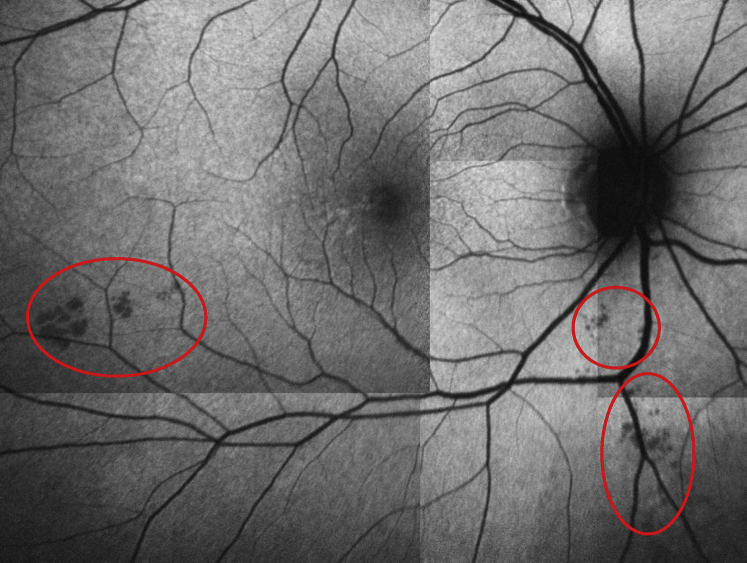
In patients with Alzheimer's disease (AD) and mild cognitive impairment, structural changes in the retina (i.e., reduced thicknesses of the ganglion cell and retinal nerve fiber layers and inclusion bodies that appear to contain beta-amyloid protein [Ab]) have been previously reported. We sought to explore whether anatomic retinal changes are detectable in the preclinical stage of AD.
A cross-sectional study (as part of an ongoing longitudinal cohort study) involving 63 cognitively normal adults, all of whom have a parent with AD and subjective memory complaints. We compared neocortical amyloid aggregation (florbetapir PET imaging) to retinal spectral domain optical coherence tomography (SD-OCT) markers of possible disease burden. Retinal biomarkers, including the number and surface area of retinal inclusion bodies and the thickness of retinal neuronal layers, were compared across groups with high vs. low neocortical beta-amyloid load.
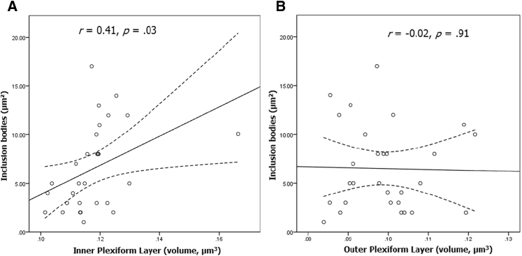
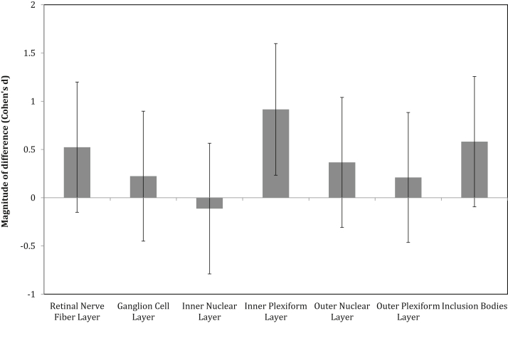
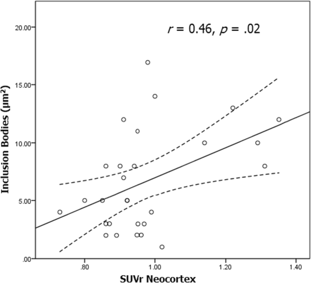
The surface area of inclusion bodies increased as a function of cortical amyloid burden. Additionally, there was a trend toward a selective volume increase in the inner plexiform layer (IPL; a layer rich in cholinergic activity) of the retina in Aβ+ relative to Aβ- participants, and IPL volume was correlated with the surface area of retinal inclusion bodies.
These initial results suggest that retinal imaging may be a potential cost-effective and noninvasive technique that can be used to identify those at-risk for AD. Layer-specific changes in the IPL and their association with surface area of inclusion bodies are discussed as a possible reflection of early inflammatory processes associated with cholinergic disruption and concurrent Ab accumulation in the neocortex.
在阿尔茨海默病(AD)和轻度认知障碍患者中,先前已有报道视网膜存在结构变化(即神经节细胞层和视网膜神经纤维层厚度减小以及出现似乎含有β-淀粉样蛋白[Ab]的包涵体)。我们试图探究在AD临床前阶段是否可检测到视网膜的解剖学变化。
一项横断面研究(作为正在进行的纵向队列研究的一部分),纳入63名认知正常的成年人,他们均有一位患AD的父母且有主观记忆主诉。我们将新皮质淀粉样蛋白聚集(氟代贝他吡PET成像)与可能的疾病负担的视网膜光谱域光学相干断层扫描(SD-OCT)标志物进行比较。比较了新皮质β-淀粉样蛋白负荷高与低的组之间的视网膜生物标志物,包括视网膜包涵体的数量和表面积以及视网膜神经元层的厚度。
包涵体的表面积随皮质淀粉样蛋白负担增加而增大。此外,相对于Aβ-参与者,Aβ+参与者的视网膜内网状层(IPL;富含胆碱能活性的一层)有选择性体积增加的趋势,且IPL体积与视网膜包涵体的表面积相关。
这些初步结果表明,视网膜成像可能是一种潜在的具有成本效益的非侵入性技术,可用于识别有AD风险的人群。IPL的层特异性变化及其与包涵体表面积的关联被讨论为可能反映了与胆碱能破坏和新皮质中同时存在的Ab积累相关的早期炎症过程。