Moghimi Sasan, Chen Rebecca, Hamzeh Nikoo, Khatibi Nassim, Lin Shan C
Farabi Eye Hospital, Tehran University of Medical Sciences, Tehran, Iran; Koret Vision Center, University of California, San Francisco Medical School, San Francisco, CA, USA.
Koret Vision Center, University of California, San Francisco Medical School, San Francisco, CA, USA.
J Curr Ophthalmol. 2016 Jul 12;28(4):170-175. doi: 10.1016/j.joco.2016.06.005. eCollection 2016 Dec.
To evaluate different mechanisms of primary angle closure (PAC) and to quantify anterior chamber (AC) parameters in different subtypes of angle closure disease using anterior segment optical coherence tomography (AS-OCT).
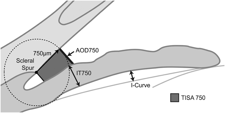
In this prospective study, 115 eyes of 115 patients with angle closure disease were included and categorized into three groups: 1) fellow eyes of acute angle closure (AAC; 40 eyes); 2) primary angle closure glaucoma (PACG; 39 eyes); and 3) primary angle closure suspect (PACS; 36 eyes). Complete ophthalmic examination including gonioscopy, A-scan biometry, and AS-OCT were performed. Based on the AS-OCT images, 4 mechanisms of PAC including pupillary block, plateau iris configuration, thick peripheral iris roll (PIR), and exaggerated lens vault were evaluated. Angle, AC, and lens parameter variables were also evaluated among the three subtypes.
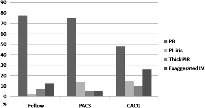
There was a statistically significant difference in the mechanism of angle closure among the three groups (p = 0.03). While the majority of fellow eyes of AAC and of PACS eyes had pupillary block mechanism (77.5% and 75%, respectively), only 48.7% of PACG eyes had dominant pupillary block mechanism (p = 0.03). The percentage of exaggerated lens vault and plateau iris mechanisms was higher in PACG eyes (25.5% and 15.4%, respectively). Fellow eyes of AAC had the shallowest AC (p = 0.01), greater iris curvature (p = 0.01), and lens vault (p = 0.02) than PACS and PACG eyes. Iris thickness was not significantly different among the three groups (p = 0.45).
Using AS-OCT, we found that there was a statistically significant difference in the underlying PAC mechanisms and quantitative AC parameters among the three subtypes of angle closure disease.
利用眼前节光学相干断层扫描(AS-OCT)评估原发性房角关闭(PAC)的不同机制,并量化房角关闭疾病不同亚型的前房(AC)参数。
在这项前瞻性研究中,纳入了115例房角关闭疾病患者的115只眼,并分为三组:1)急性房角关闭(AAC)的对侧眼(40只眼);2)原发性房角关闭型青光眼(PACG;39只眼);3)原发性房角关闭可疑者(PACS;36只眼)。进行了包括房角镜检查、A超生物测量和AS-OCT在内的完整眼科检查。基于AS-OCT图像,评估了PAC的4种机制,包括瞳孔阻滞、高原虹膜形态、厚周边虹膜卷(PIR)和晶状体拱高增大。还评估了三种亚型之间的房角、前房和晶状体参数变量。
三组之间房角关闭机制存在统计学显著差异(p = 0.03)。虽然AAC对侧眼和PACS眼的大多数具有瞳孔阻滞机制(分别为77.5%和75%),但只有48.7%的PACG眼具有主要的瞳孔阻滞机制(p = 0.03)。PACG眼中晶状体拱高增大和高原虹膜机制的比例更高(分别为25.5%和15.4%)。AAC对侧眼的前房最浅(p = 0.01),虹膜曲率大于PACS和PACG眼(p = 0.01),晶状体拱高也大于PACS和PACG眼(p = 0.02)。三组之间虹膜厚度无显著差异(p = 0.45)。
使用AS-OCT,我们发现房角关闭疾病的三种亚型在潜在的PAC机制和定量AC参数方面存在统计学显著差异。