García-Cabezas Miguel Á, John Yohan J, Barbas Helen, Zikopoulos Basilis
Neural Systems Laboratory, Department of Health Sciences, Boston University Boston, MA, USA.
Human Systems Neuroscience Laboratory, Department of Health Sciences, Boston University Boston, MA, USA.
Front Neuroanat. 2016 Nov 1;10:107. doi: 10.3389/fnana.2016.00107. eCollection 2016.
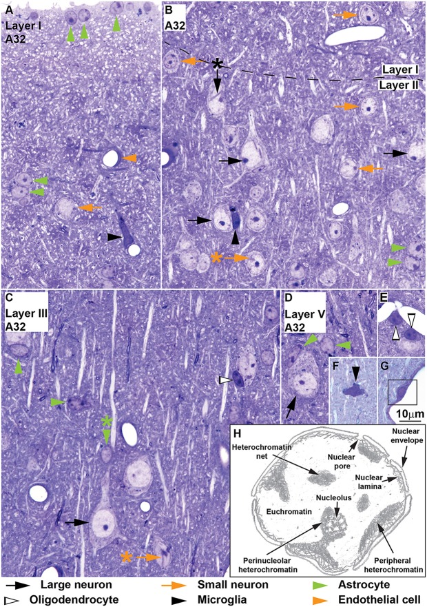
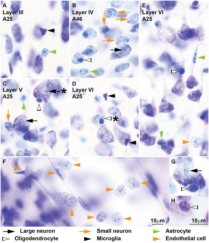
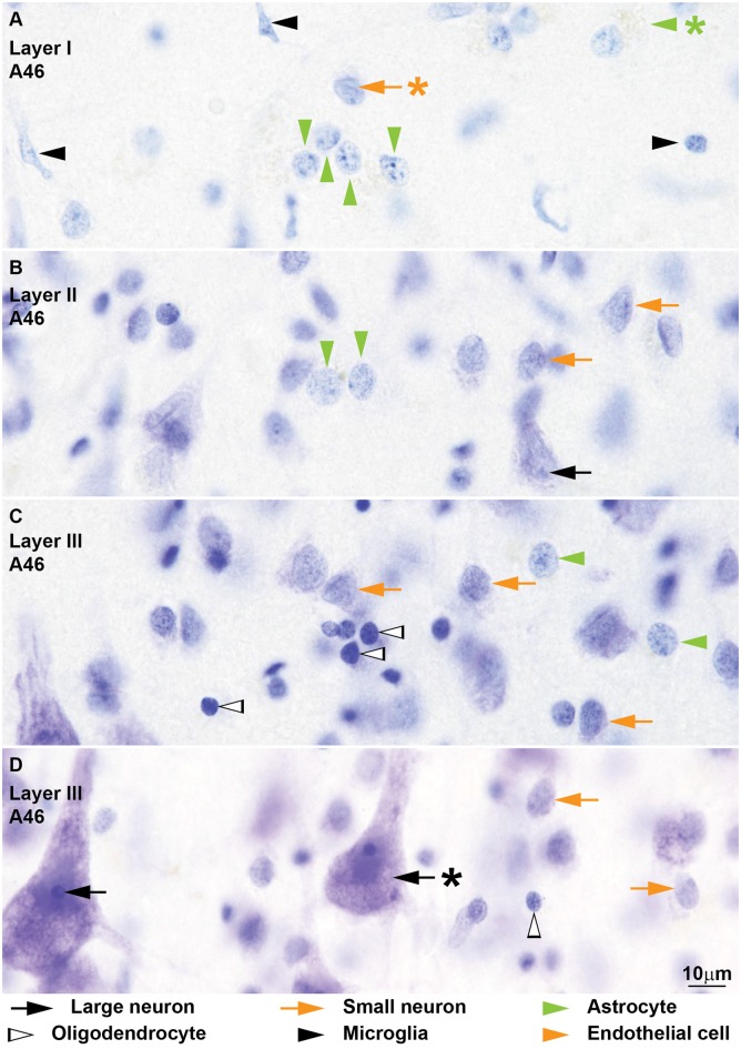
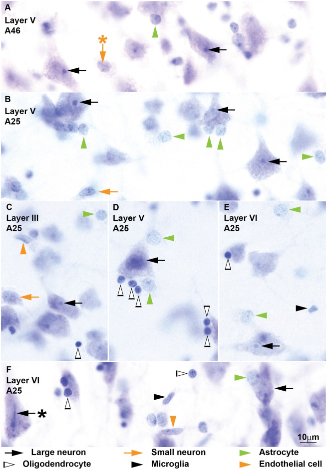
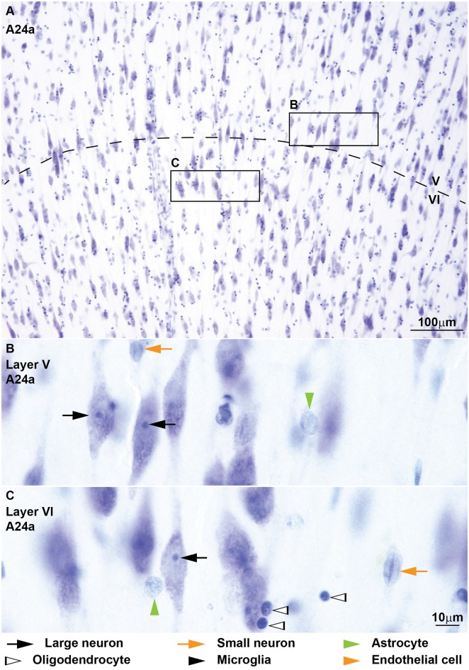
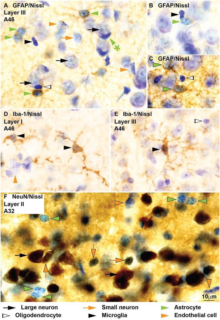
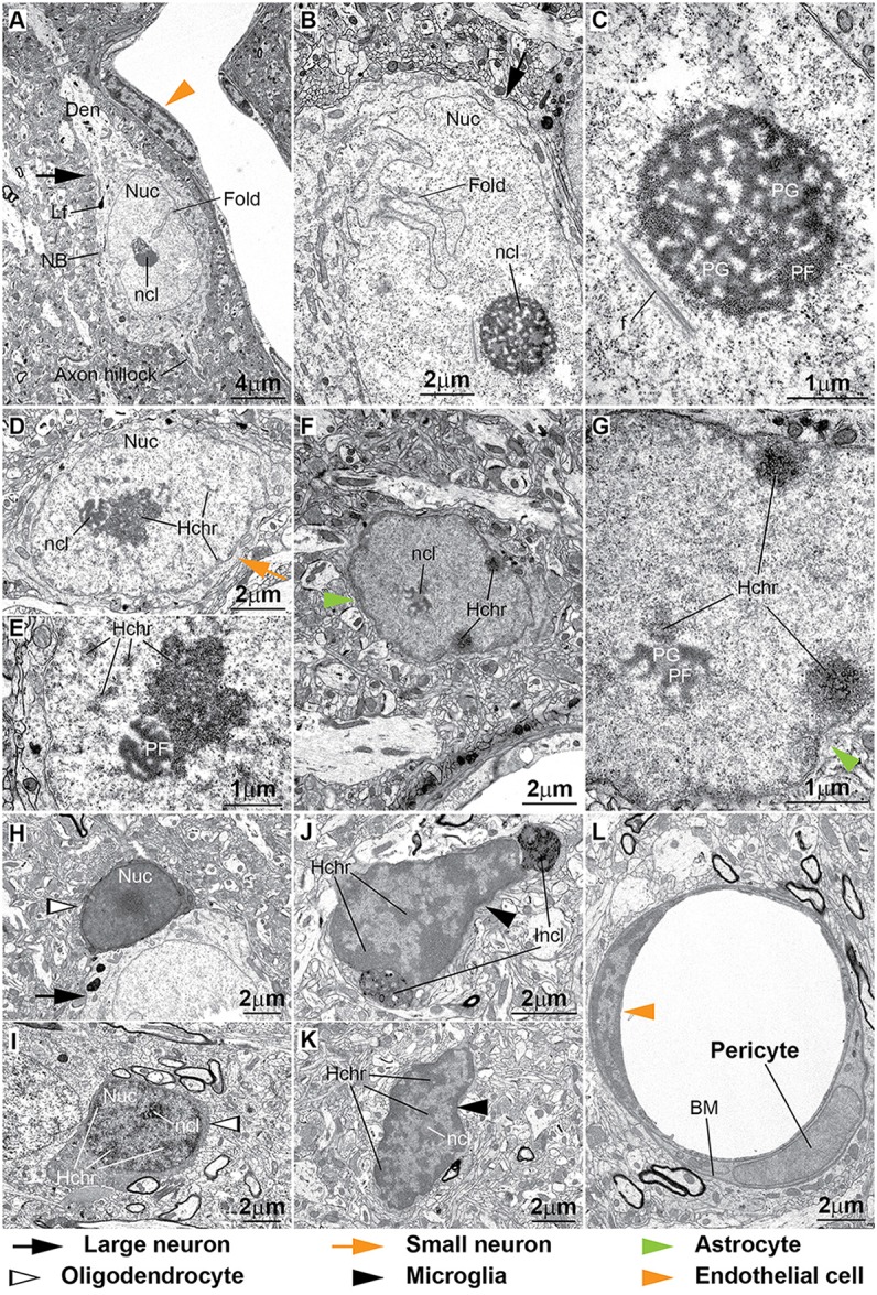
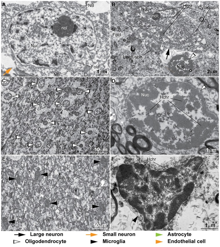
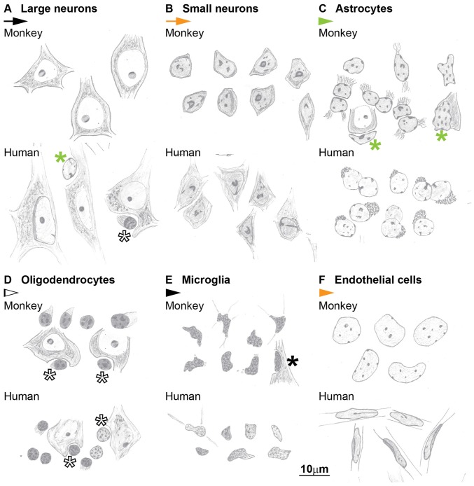
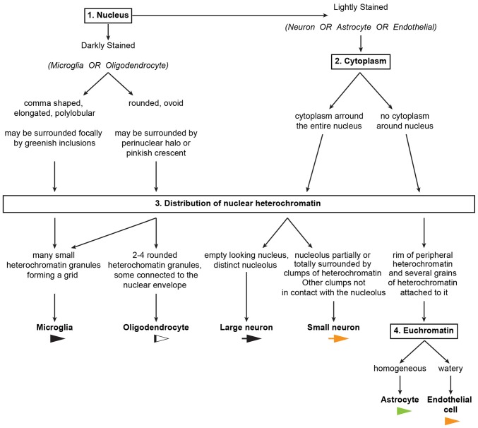
The estimation of the number or density of neurons and types of glial cells and their relative proportions in different brain areas are at the core of rigorous quantitative neuroanatomical studies. Unfortunately, the lack of detailed, updated, systematic and well-illustrated descriptions of the cytology of neurons and glial cell types, especially in the primate brain, makes such studies especially demanding, often limiting their scope and broad use. Here, following an extensive analysis of histological materials and the review of current and classical literature, we compile a list of precise morphological criteria that can facilitate and standardize identification of cells in stained sections examined under the microscope. We describe systematically and in detail the cytological features of neurons and glial cell types in the cerebral cortex of the macaque monkey and the human using semithin and thick sections stained for Nissl. We used this classical staining technique because it labels all cells in the brain in distinct ways. In addition, we corroborate key distinguishing characteristics of different cell types in sections immunolabeled for specific markers counterstained for Nissl and in ultrathin sections processed for electron microscopy. Finally, we summarize the core features that distinguish each cell type in easy-to-use tables and sketches, and structure these key features in an algorithm that can be used to systematically distinguish cellular types in the cerebral cortex. Moreover, we report high inter-observer algorithm reliability, which is a crucial test for obtaining consistent and reproducible cell counts in unbiased stereological studies. This protocol establishes a consistent framework that can be used to reliably identify and quantify cells in the cerebral cortex of primates as well as other mammalian species in health and disease.
估计不同脑区神经元的数量或密度、胶质细胞的类型及其相对比例,是严谨的定量神经解剖学研究的核心内容。不幸的是,缺乏对神经元和胶质细胞类型细胞学的详细、更新、系统且配有精美插图的描述,尤其是在灵长类动物大脑中,使得此类研究极具挑战性,常常限制了它们的范围和广泛应用。在此,在对组织学材料进行广泛分析并回顾当前及经典文献之后,我们编制了一份精确的形态学标准清单,可便于并规范在显微镜下检查的染色切片中细胞的识别。我们系统且详细地描述了猕猴和人类大脑皮质中神经元和胶质细胞类型的细胞学特征,使用的是尼氏染色的半薄切片和厚切片。我们采用这种经典染色技术是因为它能以独特方式标记大脑中的所有细胞。此外,我们在针对特定标志物进行免疫标记并以尼氏染色作为复染的切片以及用于电子显微镜检查的超薄切片中,证实了不同细胞类型的关键鉴别特征。最后,我们在便于使用的表格和示意图中总结了区分每种细胞类型的核心特征,并将这些关键特征构建成一种算法,可用于系统地区分大脑皮质中的细胞类型。此外,我们报告了观察者间算法的高可靠性,这是在无偏倚的体视学研究中获得一致且可重复细胞计数的关键测试。该方案建立了一个一致的框架,可用于可靠地识别和量化灵长类动物以及其他哺乳动物健康和疾病状态下大脑皮质中的细胞。