Hasegawa Kohei, Takaya Tomofumi, Mori Shumpei, Ito Tatsuro, Fujiwara Sei, Nishii Tatsuya, K Kono Atsushi, Shimoura Hiroyuki, Tanaka Hidekazu, Hirata Ken-Ichi
Division of Cardiovascular Medicine, Department of Internal Medicine, Kobe University Graduate School of Medicine, Japan.
Intern Med. 2016;55(22):3279-3283. doi: 10.2169/internalmedicine.55.7193. Epub 2016 Nov 15.
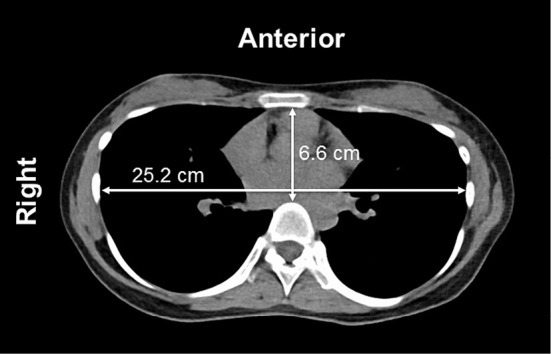
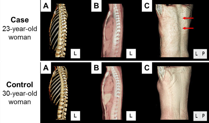
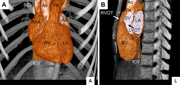
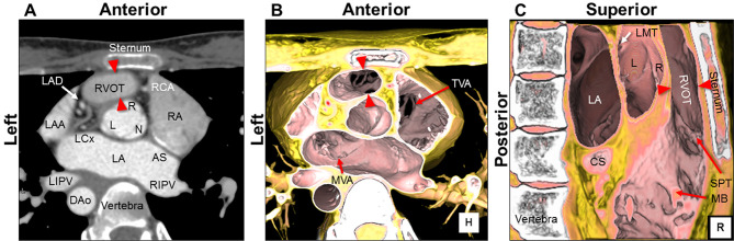
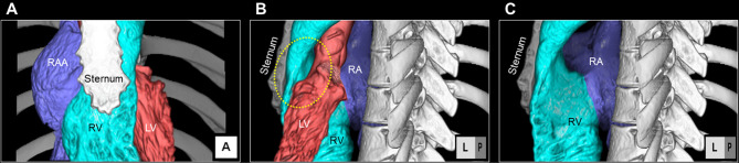
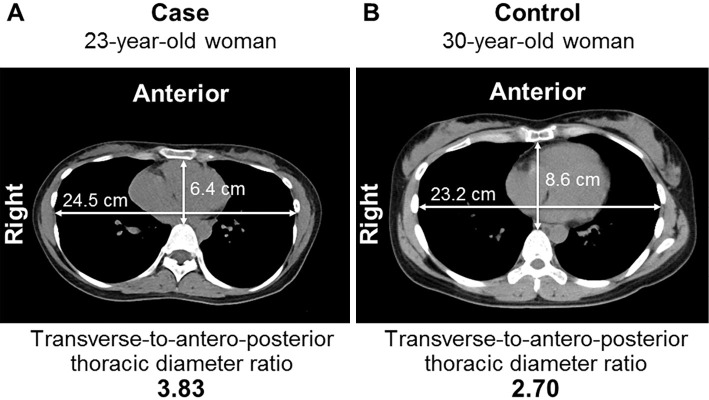
A 23-year-old asymptomatic woman was referred to our hospital for further examination of a systolic ejection murmur with fixed splitting of the second heart sound auscultated at the third left sternal border. Initial echocardiography could not detect the cause. Subsequently performed low-dose computed tomography, however, ruled out the possibility of any congenital heart diseases, but revealed a markedly shortened anteroposterior diameter of the chest, which led us to a diagnosis of straight back syndrome. A vertically oriented "pancake" appearance of the heart, straight vertebral column, and compression of the right ventricular outflow tract were clearly demonstrated on the reconstructed images.
一名23岁无症状女性因在左胸骨缘第三肋间听诊发现收缩期喷射性杂音伴第二心音固定分裂而被转诊至我院进一步检查。初次超声心动图检查未发现病因。然而,随后进行的低剂量计算机断层扫描排除了任何先天性心脏病的可能性,但显示胸部前后径明显缩短,这使我们诊断为直背综合征。重建图像清晰显示心脏呈垂直的“煎饼”样外观、脊柱变直以及右心室流出道受压。