Tretter Justin T, Gupta Saurabh Kumar, Izawa Yu, Nishii Tatsuya, Mori Shumpei
Heart Institute, Cincinnati Children's Hospital Medical Center, Cincinnati, OH 45229, USA.
Department of Pediatrics, University of Cincinnati College of Medicine, Cincinnati, OH 45229, USA.
J Cardiovasc Dev Dis. 2020 Aug 12;7(3):30. doi: 10.3390/jcdd7030030.
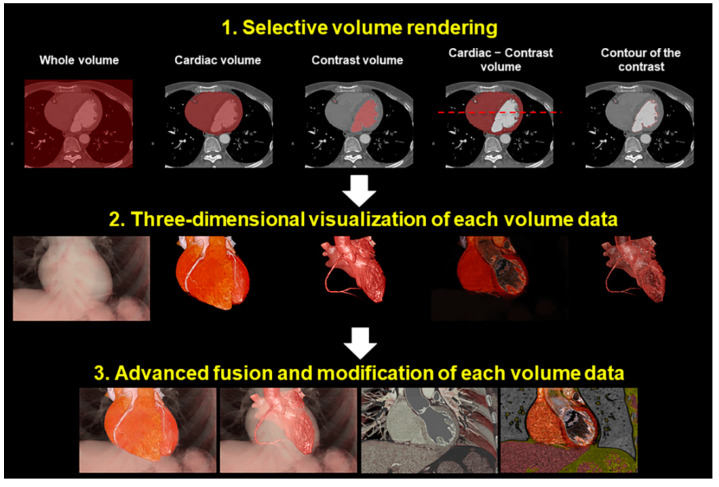
Traditionally, gross cardiac anatomy has been described mainly based on the findings in the dissection suite. Analyses of heart specimens have contributed immensely towards building a fundamental knowledge of cardiac anatomy. However, there are limitations in analyzing the autopsied heart removed from the thorax. Three-dimensional imaging allows visualization of the blood-filled heart in vivo in attitudinally appropriate fashion. This is of paramount importance for not only demonstration of cardiac anatomy for educational purposes, but also for the detailed anatomical evaluation in patients with acquired and congenital heart disease. In this review, we discuss the advantages of three-dimensional imaging, specifically focusing on virtual dissection, a volume rendering-based reconstruction technique using computed tomographic data. We highlight examples of three-dimensional imaging in both education and guiding patient management.
传统上,心脏大体解剖主要是基于解剖室的发现来描述的。对心脏标本的分析为构建心脏解剖学的基础知识做出了巨大贡献。然而,分析从胸腔取出的尸检心脏存在局限性。三维成像能够以符合体位要求的方式在体内可视化充满血液的心脏。这不仅对于出于教育目的展示心脏解剖结构至关重要,而且对于后天性和先天性心脏病患者的详细解剖评估也至关重要。在本综述中,我们讨论三维成像的优势,特别关注虚拟解剖,这是一种使用计算机断层扫描数据的基于容积再现的重建技术。我们重点介绍三维成像在教育和指导患者管理方面的实例。