Baldivia Sarah, Levy Alexander, Hegde Shylaja, Aper Stijn J A, Merkx Maarten, Grytz Rafael
Department of Biomedical Engineering, University of Alabama at Birmingham, Birmingham, Alabama, United States of America.
Department of Vision Sciences, University of Alabama at Birmingham, Birmingham, Alabama, United States of America.
PLoS One. 2016 Nov 21;11(11):e0166644. doi: 10.1371/journal.pone.0166644. eCollection 2016.
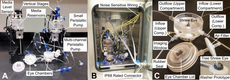
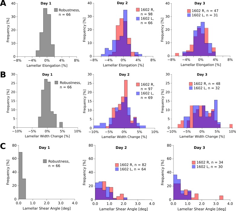
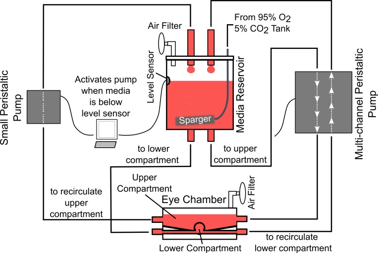
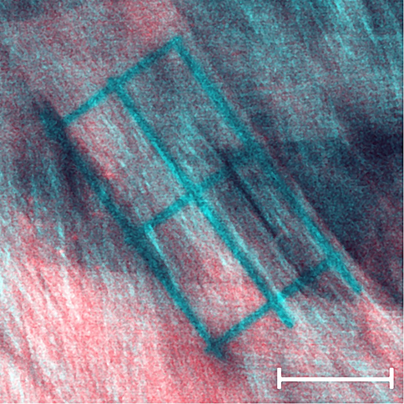
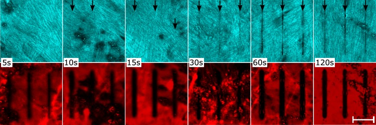
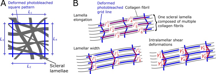
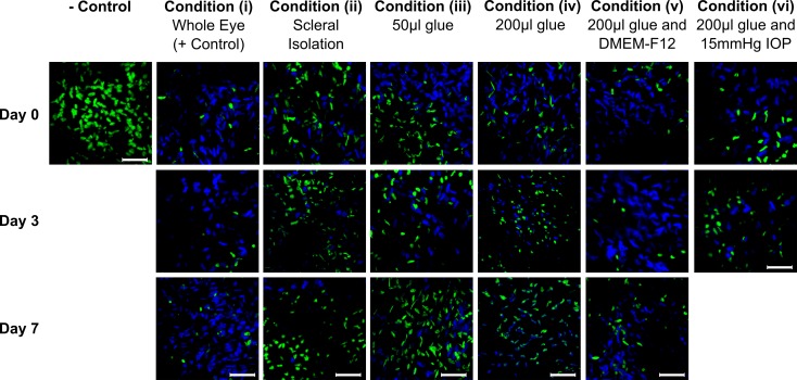
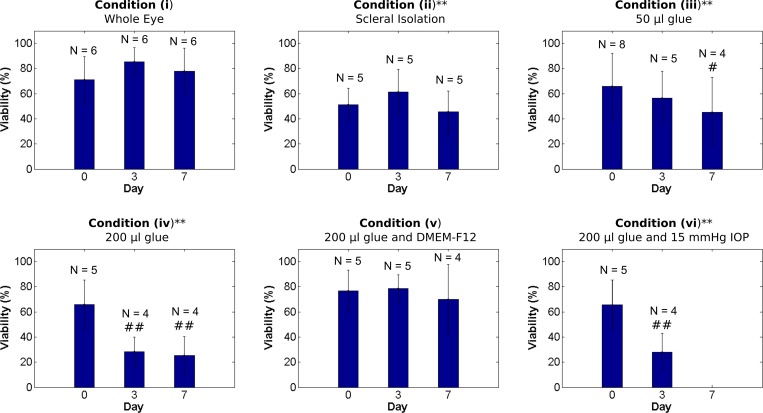
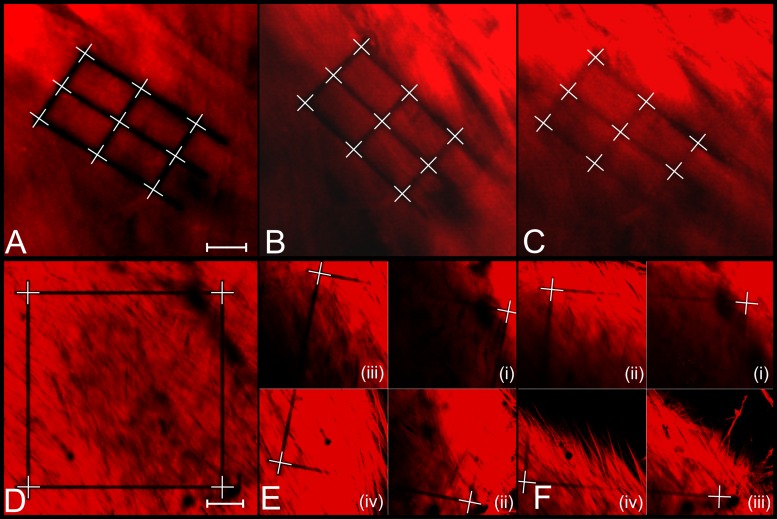
Increasing evidence suggests that unknown collagen remodeling mechanisms in the sclera underlie myopia development. We are proposing a novel organ culture system in combination with two-photon fluorescence imaging to quantify collagen remodeling at the tissue- and lamella-level. Tree shrew scleral shells were cultured up to 7 days in serum-free media and cellular viability was investigated under: (i) minimal tissue manipulations; (ii) removal of intraocular tissues; gluing the eye to a washer using (iii) 50 μL and (iv) 200 μL of cyanoacrylate adhesive; (v) supplementing media with Ham's F-12 Nutrient Mixture; and (vi) culturing eyes subjected to 15 mmHg intraocular pressure in our new bioreactor. Two scleral shells of normal juvenile tree shrews were fluorescently labeled using a collagen specific protein and cultured in our bioreactor. Using two-photon microscopy, grid patterns were photobleached into and across multiple scleral lamellae. These patterns were imaged daily for 3 days, and tissue-/lamella-level strains were calculated from the deformed patterns. No significant reduction in cell viability was observed under conditions (i) and (v). Compared to condition (i), cell viability was significantly reduced starting at day 0 (condition (ii)) and day 3 (conditions (iii, iv, vi)). Tissue-level strain and intralamellar shear angel increased significantly during the culture period. Some scleral lamellae elongated while others shortened. Findings suggest that tree shrew sclera can be cultured in serum-free media for 7 days with no significant reduction in cell viability. Scleral fibroblasts are sensitive to tissue manipulations and tissue gluing. However, Ham's F-12 Nutrient Mixture has a protective effect on cell viability and can offset the cytotoxic effect of cyanoacrylate adhesive. This is the first study to quantify collagen micro-deformations over a prolonged period in organ culture providing a new methodology to study scleral remodeling in myopia.
越来越多的证据表明,巩膜中未知的胶原蛋白重塑机制是近视发展的基础。我们提出了一种结合双光子荧光成像的新型器官培养系统,以在组织和板层水平上量化胶原蛋白重塑。树鼩巩膜壳在无血清培养基中培养长达7天,并在以下条件下研究细胞活力:(i)最小限度的组织操作;(ii)去除眼内组织;使用(iii)50μL和(iv)200μL氰基丙烯酸酯粘合剂将眼睛粘在垫圈上;(v)在培养基中补充哈姆氏F-12营养混合物;以及(vi)在我们的新型生物反应器中培养承受15 mmHg眼内压的眼睛。使用胶原蛋白特异性蛋白对正常幼年树鼩的两个巩膜壳进行荧光标记,并在我们的生物反应器中培养。使用双光子显微镜,将网格图案光漂白到多个巩膜板层并穿过这些板层。这些图案每天成像3天,并根据变形图案计算组织/板层水平的应变。在条件(i)和(v)下未观察到细胞活力的显著降低。与条件(i)相比,从第0天(条件(ii))和第3天(条件(iii、iv、vi))开始,细胞活力显著降低。在培养期间,组织水平应变和板层内剪切角显著增加。一些巩膜板层伸长而另一些缩短。研究结果表明,树鼩巩膜可以在无血清培养基中培养7天,而细胞活力没有显著降低。巩膜成纤维细胞对组织操作和组织粘贴敏感。然而,哈姆氏F-12营养混合物对细胞活力有保护作用,可以抵消氰基丙烯酸酯粘合剂的细胞毒性作用。这是第一项在器官培养中长时间量化胶原蛋白微变形的研究,并提供了一种研究近视中巩膜重塑的新方法。