Henriques Paulo S G, Okajima Luciana S, Nunes Marcelo P, Montalli Victor A M
Department of Periodontology, São Leopoldo Mandic Institute and Research Center, Campinas, SP, Brazil.
Department of Oral Pathology, São Leopoldo Mandic Institute and Research Center, Campinas, SP, Brazil.
Case Rep Dent. 2016;2016:6874235. doi: 10.1155/2016/6874235. Epub 2016 Nov 7.
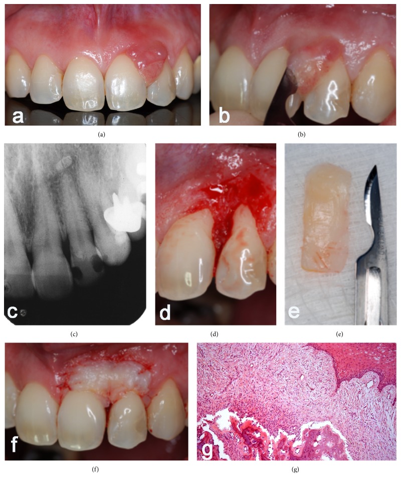
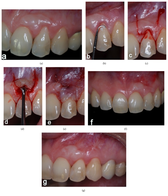
When lesions in soft tissue reach the gingival margin, they can produce aesthetic defects during its permanence and after its removal. Periodontal plastic surgery allows the correction of the gingival contour using different techniques. This paper is a case report of a peripheral ossifying fibroma removal in the interproximal area of teeth 21 and 22 in addition to root coverage of the affected area through two surgical phases: keratinized gingival tissue augmentation surgery with free gingival graft concurrent with removal of the lesion and, in a second stage, root coverage by performing coronally advanced flap technique with a follow-up of five years. The initial results achieved, which were root coverage of 100% after 6 months, promoted an adequate gingival contour and prevented the development of a mucogingival defect or a root exposure with its functional and aesthetic consequences. After five years, the results showed long term success of the techniques, where the margin remained stable with complete root coverage and tissues were stable and harmonic in color.
当软组织病变累及牙龈边缘时,在病变持续存在期间及其切除后均会产生美观缺陷。牙周整形手术可采用不同技术矫正牙龈外形。本文是一例在21和22牙邻间隙切除外周骨化性纤维瘤的病例报告,同时通过两个手术阶段对患区进行牙根覆盖:在切除病变的同时,采用游离龈瓣移植术进行角化龈组织增量手术,第二阶段采用冠向推进瓣技术进行牙根覆盖,并进行了五年的随访。最初取得的结果是6个月后牙根覆盖率达到100%,促进了牙龈外形的正常化,并防止了膜龈缺损或牙根暴露及其功能和美观后果的发生。五年后,结果显示这些技术取得了长期成功,边缘保持稳定,牙根完全覆盖,组织稳定且颜色协调。