• 文献检索
  • 文档翻译
  • 深度研究
  • 学术资讯
  • Suppr Zotero 插件Zotero 插件
  • 邀请有礼
  • 套餐&价格
  • 历史记录
应用&插件
Suppr Zotero 插件Zotero 插件浏览器插件Mac 客户端Windows 客户端微信小程序
定价
高级版会员购买积分包购买API积分包
服务
文献检索文档翻译深度研究API 文档MCP 服务
关于我们
关于 Suppr公司介绍联系我们用户协议隐私条款
关注我们

Suppr 超能文献

核心技术专利:CN118964589B侵权必究
粤ICP备2023148730 号-1Suppr @ 2026

文献检索

告别复杂PubMed语法,用中文像聊天一样搜索,搜遍4000万医学文献。AI智能推荐,让科研检索更轻松。

立即免费搜索

文件翻译

保留排版,准确专业,支持PDF/Word/PPT等文件格式,支持 12+语言互译。

免费翻译文档

深度研究

AI帮你快速写综述,25分钟生成高质量综述,智能提取关键信息,辅助科研写作。

立即免费体验

复发缓解型多发性硬化症在振幅配置下的功能性皮质变化:一项静息态功能磁共振成像研究

Functional cortical changes in relapsing-remitting multiple sclerosis at amplitude configuration: a resting-state fMRI study.

作者信息

Liu Heng, Chen Hua, Wu Bo, Zhang Tijiang, Wang Jinhui, Huang Kexin, Song Ganjun, Zhan Jian

机构信息

Department of Radiology, Affiliated Hospital of Zunyi Medical University, Medical Imaging Center of Guizhou Province, Zunyi, Guizhou.

Department of Psychology, Hangzhou Normal University; Zhejiang Key Laboratory for Research in Assessment of Cognitive Impairments, Hangzhou.

出版信息

Neuropsychiatr Dis Treat. 2016 Nov 25;12:3031-3039. doi: 10.2147/NDT.S120909. eCollection 2016.

DOI:10.2147/NDT.S120909
PMID:27932883
原文链接:https://pmc.ncbi.nlm.nih.gov/articles/PMC5135476/
Abstract

OBJECTIVE

The aim of this study was to explore the amplitude of spontaneous brain activity fluctuations in patients with relapsing-remitting multiple sclerosis (RRMS) using the amplitude of low-frequency fluctuation (ALFF) method.

METHODS

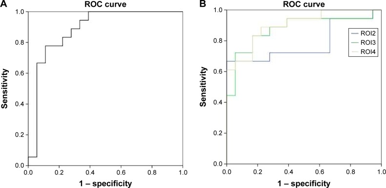
ALFF and SPM8 were utilized to assess alterations in regional spontaneous brain activities in patients with RRMS in comparison with healthy controls (HCs). The beta values of altered brain regions between patients with RRMS and HCs were extracted, and a receiver operating characteristic (ROC) curve was generated to calculate the sensitivities and specificities of these different brain areas for distinguishing patients with RRMS from HCs. Pearson correlation analyses were applied to assess the relationships between the beta values of altered brain regions and disease duration and Expanded Disability Status Scale (EDSS) score.

PATIENTS AND PARTICIPANTS

A total of 18 patients with RRMS (13 females; five males) and 18 sex-, age-, and education-matched HCs (14 females; four males) were recruited for this study.

MEASUREMENTS AND RESULTS

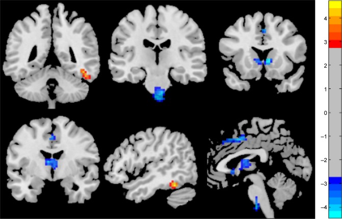
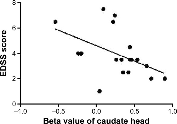
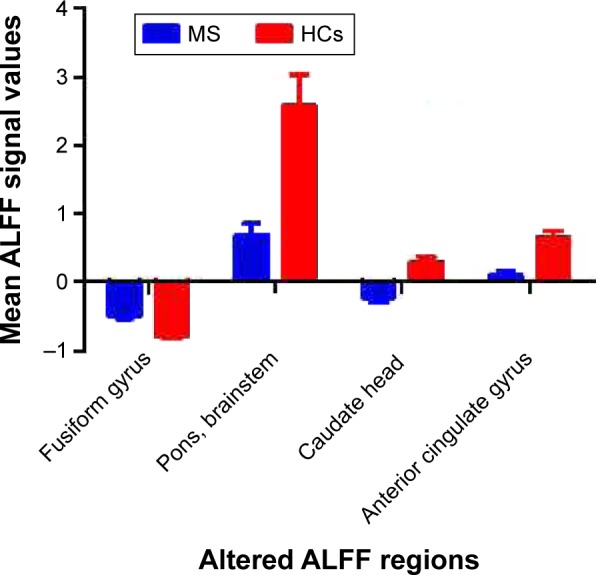
Compared with HCs, patients with RRMS showed higher ALFF responses in the right fusiform gyrus (Brodmann area [BA] 37) and lower ALFF responses in the bilateral anterior cingulate cortices (BA 24 and 32), bilateral heads of the caudate nuclei, and bilateral brainstem. The ROC analysis revealed that the beta values of these abnormal brain areas showed high degrees of sensitivity and specificity for distinguishing patients with RRMS from HCs. The EDSS score showed a significant negative Pearson correlation with the beta value of the caudate head (=-0.474, =0.047).

CONCLUSION

RRMS is associated with disturbances in spontaneous regional brain activity in specific areas, and these specific abnormalities may provide important information about the neural mechanisms underlying behavioral impairment in RRMS.

摘要

目的

本研究旨在采用低频振幅(ALFF)方法探究复发缓解型多发性硬化症(RRMS)患者脑自发活动波动的幅度。

方法

与健康对照者(HCs)相比,利用ALFF和SPM8评估RRMS患者局部脑自发活动的改变。提取RRMS患者和HCs之间脑区改变的β值,并生成受试者工作特征(ROC)曲线,以计算这些不同脑区区分RRMS患者和HCs的敏感性和特异性。应用Pearson相关性分析评估脑区改变的β值与病程及扩展残疾状态量表(EDSS)评分之间的关系。

患者与参与者

本研究共招募了18例RRMS患者(13例女性;5例男性)和18例性别、年龄及教育程度匹配的HCs(14例女性;4例男性)。

测量与结果

与HCs相比,RRMS患者右侧梭状回(布罗德曼区[BA]37)的ALFF反应较高,而双侧前扣带回皮质(BA 24和32)、双侧尾状核头部及双侧脑干的ALFF反应较低。ROC分析显示,这些异常脑区的β值在区分RRMS患者和HCs方面具有高度的敏感性和特异性。EDSS评分与尾状核头部的β值呈显著负Pearson相关性(r = -0.474,P = 0.047)。

结论

RRMS与特定区域的脑自发活动紊乱有关,这些特定异常可能为RRMS行为障碍的神经机制提供重要信息。

https://cdn.ncbi.nlm.nih.gov/pmc/blobs/6b74/5135476/a80165bef4ed/ndt-12-3031Fig4.jpg
https://cdn.ncbi.nlm.nih.gov/pmc/blobs/6b74/5135476/4df51406216d/ndt-12-3031Fig1.jpg
https://cdn.ncbi.nlm.nih.gov/pmc/blobs/6b74/5135476/db9252568612/ndt-12-3031Fig2.jpg
https://cdn.ncbi.nlm.nih.gov/pmc/blobs/6b74/5135476/649aabbf7ec0/ndt-12-3031Fig3.jpg
https://cdn.ncbi.nlm.nih.gov/pmc/blobs/6b74/5135476/a80165bef4ed/ndt-12-3031Fig4.jpg

相似文献

1
Functional cortical changes in relapsing-remitting multiple sclerosis at amplitude configuration: a resting-state fMRI study.复发缓解型多发性硬化症在振幅配置下的功能性皮质变化:一项静息态功能磁共振成像研究
Neuropsychiatr Dis Treat. 2016 Nov 25;12:3031-3039. doi: 10.2147/NDT.S120909. eCollection 2016.
2
Relapsing-Remitting Multiple Sclerosis Is Associated With Regional Brain Activity Deficits in Motor- and Cognitive-Related Brain Areas.复发缓解型多发性硬化症与运动和认知相关脑区的局部脑活动缺陷有关。
Front Neurol. 2019 Nov 26;10:1136. doi: 10.3389/fneur.2019.01136. eCollection 2019.
3
Altered spontaneous brain activity patterns in patients with retinal vein occlusion indicated by the amplitude of low-frequency fluctuation: A functional magnetic resonance imaging study.基于低频振幅波动的视网膜静脉阻塞患者自发脑活动模式改变:一项功能磁共振成像研究
Exp Ther Med. 2019 Sep;18(3):2063-2071. doi: 10.3892/etm.2019.7770. Epub 2019 Jul 12.
4
Brain plasticity in relapsing-remitting multiple sclerosis: evidence from resting-state fMRI.复发缓解型多发性硬化症中的大脑可塑性:静息态 fMRI 的证据。
J Neurol Sci. 2011 May 15;304(1-2):127-31. doi: 10.1016/j.jns.2011.01.023. Epub 2011 Feb 23.
5
Increased thalamic intrinsic oscillation amplitude in relapsing-remitting multiple sclerosis associated with the slowed cognitive processing.复发缓解型多发性硬化症中丘脑固有振荡幅度增加与认知加工减慢有关。
Clin Imaging. 2014 Sep-Oct;38(5):605-10. doi: 10.1016/j.clinimag.2014.05.006. Epub 2014 May 21.
6
Altered spontaneous brain activity patterns in patients with corneal ulcer using amplitude of low-frequency fluctuation: An fMRI study.基于低频振幅波动的角膜溃疡患者自发脑活动模式改变:一项功能磁共振成像研究
Exp Ther Med. 2019 Jul;18(1):125-132. doi: 10.3892/etm.2019.7550. Epub 2019 May 7.
7
Investigating amplitude of low-frequency fluctuation and possible links with cognitive impairment in childhood and adolescence onset schizophrenia: a correlation study.探究儿童和青少年期起病精神分裂症低频波动幅度及其与认知障碍的潜在联系:一项相关性研究。
Front Psychiatry. 2024 Feb 15;15:1288955. doi: 10.3389/fpsyt.2024.1288955. eCollection 2024.
8
Altered spontaneous brain activity patterns in patients with neovascular glaucoma using amplitude of low-frequency fluctuations: A functional magnetic resonance imaging study.利用低频波动幅度研究新生血管性青光眼患者自发脑活动模式的改变:一项功能磁共振成像研究。
Brain Behav. 2021 Mar;11(3):e02018. doi: 10.1002/brb3.2018. Epub 2021 Jan 1.
9
Altered intrinsic brain activities in patients with acute eye pain using amplitude of low-frequency fluctuation: a resting-state fMRI study.基于低频振幅波动的急性眼痛患者脑内固有活动改变:一项静息态功能磁共振成像研究
Neuropsychiatr Dis Treat. 2018 Jan 9;14:251-257. doi: 10.2147/NDT.S150051. eCollection 2018.
10
Altered intra- and interregional synchronization in relapsing-remitting multiple sclerosis: a resting-state fMRI study.复发缓解型多发性硬化症中区域内和区域间同步性的改变:一项静息态功能磁共振成像研究
Neuropsychiatr Dis Treat. 2016 Apr 15;12:853-62. doi: 10.2147/NDT.S98962. eCollection 2016.

引用本文的文献

1
Altered intrinsic brain activity in patients with CSF1R-related leukoencephalopathy.CSF1R 相关脑白质病患者的脑内固有活动改变。
Brain Imaging Behav. 2022 Aug;16(4):1842-1853. doi: 10.1007/s11682-022-00646-5. Epub 2022 Apr 7.
2
Resting-state functional MRI demonstrates brain network reorganization in neuromyelitis optica spectrum disorder (NMOSD).静息态功能磁共振成像显示视神经脊髓炎谱系疾病(NMOSD)中的脑网络重组。
PLoS One. 2019 Jan 29;14(1):e0211465. doi: 10.1371/journal.pone.0211465. eCollection 2019.
3
The relationship between amplitude of low frequency fluctuations and gray matter volume of the mirror neuron system: Differences between low disability multiple sclerosis patients and healthy controls.

本文引用的文献

1
Functional Magnetic Resonance Imaging with Concurrent Urodynamic Testing Identifies Brain Structures Involved in Micturition Cycle in Patients with Multiple Sclerosis.功能磁共振成像联合尿动力学检查识别多发性硬化症患者排尿周期相关脑区。
J Urol. 2017 Feb;197(2):438-444. doi: 10.1016/j.juro.2016.09.077. Epub 2016 Sep 21.
2
Novel fMRI working memory paradigm accurately detects cognitive impairment in multiple sclerosis.新型功能磁共振成像工作记忆范式可准确检测多发性硬化症中的认知障碍。
Mult Scler. 2017 May;23(6):836-847. doi: 10.1177/1352458516666186. Epub 2016 Sep 9.
3
Functional connectivity of paired default mode network subregions in primary insomnia.
低频波动幅度与镜像神经元系统灰质体积之间的关系:低残疾多发性硬化症患者与健康对照者的差异。
IBRO Rep. 2018 Sep 27;5:60-66. doi: 10.1016/j.ibror.2018.09.002. eCollection 2018 Dec.
原发性失眠中默认模式网络配对子区域的功能连接性
Neuropsychiatr Dis Treat. 2015 Dec 16;11:3085-93. doi: 10.2147/NDT.S95224. eCollection 2015.
4
Gender Differences in Regional Brain Activity in Patients with Chronic Primary Insomnia: Evidence from a Resting-State fMRI Study.慢性原发性失眠患者脑区活动的性别差异:一项静息态功能磁共振成像研究的证据
J Clin Sleep Med. 2016 Mar;12(3):363-74. doi: 10.5664/jcsm.5586.
5
Abnormal Baseline Brain Activity in Patients With Multiple Sclerosis With Simple Spinal Cord Involvement Detected by Resting-State Functional Magnetic Resonance Imaging.静息态功能磁共振成像检测发现单纯脊髓受累的多发性硬化症患者基线脑活动异常
J Comput Assist Tomogr. 2015 Nov-Dec;39(6):866-75. doi: 10.1097/RCT.0000000000000299.
6
Disturbed spontaneous brain activity pattern in patients with primary angle-closure glaucoma using amplitude of low-frequency fluctuation: a fMRI study.利用低频振幅波动对原发性闭角型青光眼患者自发性脑活动模式的干扰:一项功能磁共振成像研究
Neuropsychiatr Dis Treat. 2015 Jul 29;11:1877-83. doi: 10.2147/NDT.S87596. eCollection 2015.
7
Abnormal cerebellar functional MRI connectivity in patients with paediatric multiple sclerosis.小儿多发性硬化症患者小脑功能磁共振成像连接异常。
Mult Scler. 2016 Mar;22(3):292-301. doi: 10.1177/1352458515592191. Epub 2015 Jul 10.
8
Multimodal Quantitative MR Imaging of the Thalamus in Multiple Sclerosis and Neuromyelitis Optica.多模态定量磁共振成像在多发性硬化症和视神经脊髓炎中的丘脑研究。
Radiology. 2015 Dec;277(3):784-92. doi: 10.1148/radiol.2015142786. Epub 2015 Jun 4.
9
Cardiorespiratory fitness and its association with thalamic, hippocampal, and basal ganglia volumes in multiple sclerosis.心血管呼吸适能及其与多发性硬化症中丘脑、海马体和基底神经节体积的关联。
Neuroimage Clin. 2015 Feb 26;7:661-6. doi: 10.1016/j.nicl.2015.02.017. eCollection 2015.
10
Long-term total sleep deprivation decreases the default spontaneous activity and connectivity pattern in healthy male subjects: a resting-state fMRI study.长期完全睡眠剥夺降低健康男性被试的默认自发活动和连接模式:一项静息态 fMRI 研究。
Neuropsychiatr Dis Treat. 2015 Mar 19;11:761-72. doi: 10.2147/NDT.S78335. eCollection 2015.