Diabetes Research Institute, Metabolism, Nutrigenomics and Cellular Differentiation Unit, San Raffaele Scientific Institute, 60 Olgettina street, 20132, Milan, Italy.
Department of Biomedical Sciences for Health, University of Milan, Milan, Italy.
Endocrine. 2017 Oct;58(1):33-45. doi: 10.1007/s12020-016-1181-5. Epub 2016 Dec 8.
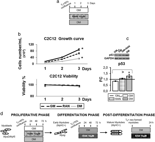
The purpose of this study is to investigate Ranolazine action on skeletal muscle differentiation and mitochondrial oxidative phenomena. Ranolazine, an antianginal drug, which acts blocking the late INaL current, was shown to lower hemoglobin A1c in patients with diabetes. In the present study, we hypothesized an action of Ranolazine on skeletal muscle cells regeneration and oxidative process, leading to a reduction of insulin resistance.
10 μM Ranolazine was added to C2C12 murine myoblastic cells during proliferation, differentiation and newly formed myotubes.
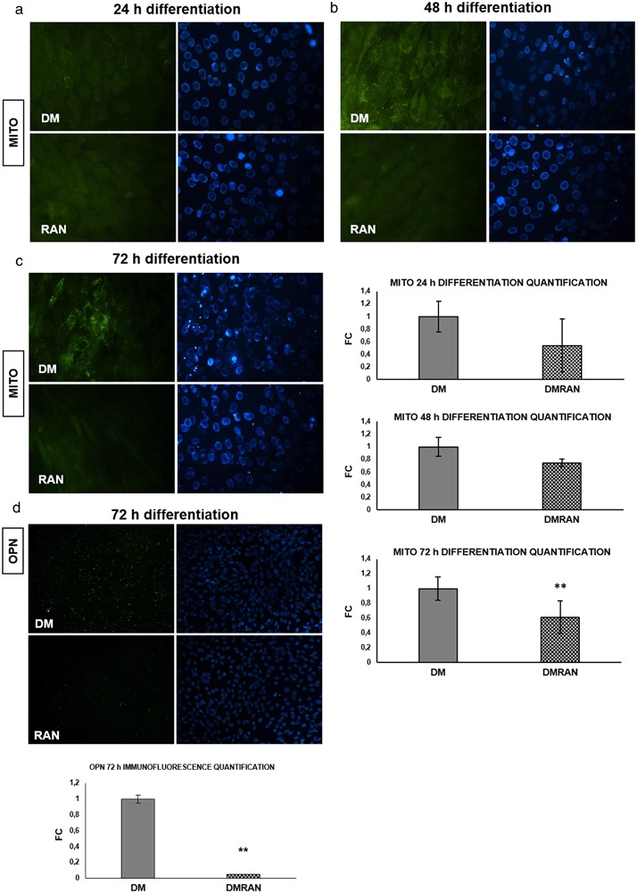
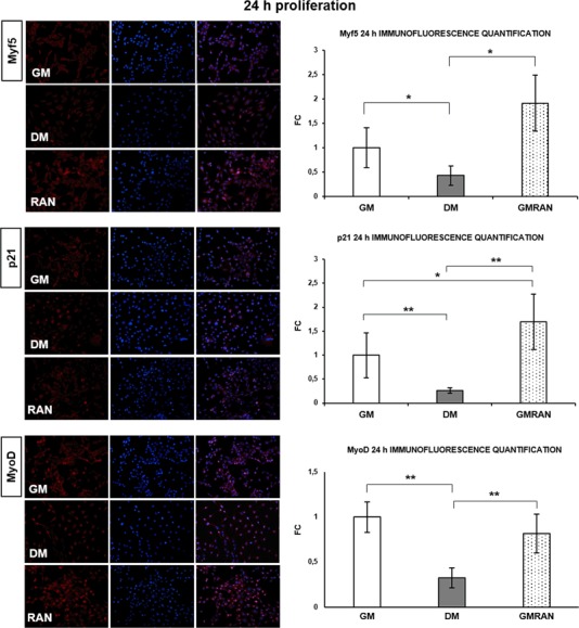
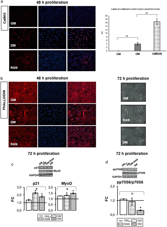
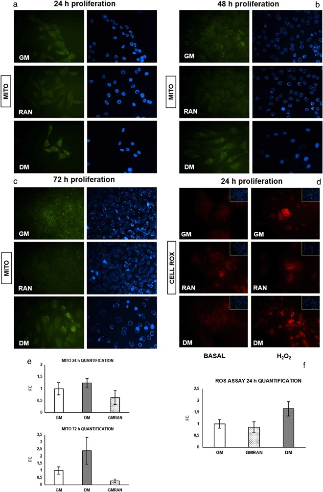
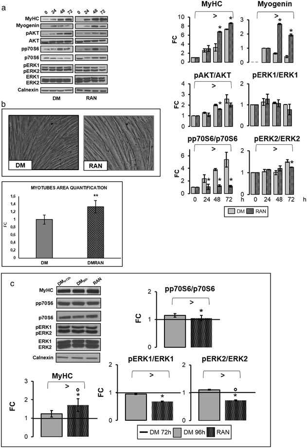
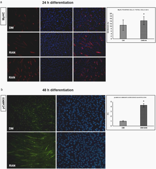
Ranolazine promoted the development of a specific myogenic phenotype: increasing the expression of myogenic regulator factors and inhibiting cell cycle progression factor (p21). Ranolazine stimulated calcium signaling (calmodulin-dependent kinases) and reduced reactive oxygen species levels. Furthermore, Ranolazine maintained mitochondrial homeostasis. During the differentiation phase, Ranolazine promoted myotubes formation. Ranolazine did not modify kinases involved in skeletal muscle differentiation and glucose uptake (extracellular signal-regulated kinases 1/2 and AKT pathways), but activated calcium signaling pathways. During proliferation, Ranolazine did not modify the number of mitochondria while decreasing osteopontin protein levels. Lastly, neo-formed myotubes treated with Ranolazine showed typical hypertrophic phenotype.
In conclusion, our results indicate that Ranolazine stimulates myogenesis and reduces a pro-oxidant inflammation/oxidative condition, activating a calcium signaling pathway. These newly described mechanisms may partially explain the glucose lowering effect of the drug.
本研究旨在探讨雷诺嗪对骨骼肌分化和线粒体氧化现象的作用。雷诺嗪是一种抗心绞痛药物,可阻断晚期 INaL 电流,有研究表明其可降低糖尿病患者的血红蛋白 A1c。在本研究中,我们假设雷诺嗪对骨骼肌细胞再生和氧化过程有作用,从而降低胰岛素抵抗。
在增殖、分化和新形成的肌管阶段,向 C2C12 鼠成肌细胞中添加 10 μM 雷诺嗪。
雷诺嗪促进了特定的成肌表型的发展:增加肌生成调节因子的表达,抑制细胞周期进程因子(p21)。雷诺嗪刺激钙信号(钙调蛋白依赖性激酶)并降低活性氧水平。此外,雷诺嗪维持线粒体稳态。在分化阶段,雷诺嗪促进肌管形成。雷诺嗪不改变参与骨骼肌分化和葡萄糖摄取的激酶(细胞外信号调节激酶 1/2 和 AKT 途径),但激活了钙信号通路。在增殖阶段,雷诺嗪不改变线粒体的数量,同时降低骨桥蛋白的蛋白水平。最后,用雷诺嗪处理的新形成的肌管表现出典型的肥大表型。
总之,我们的结果表明,雷诺嗪刺激肌生成并降低促氧化炎症/氧化状态,激活钙信号通路。这些新描述的机制可能部分解释了该药物的降血糖作用。