• 文献检索
  • 文档翻译
  • 深度研究
  • 学术资讯
  • Suppr Zotero 插件Zotero 插件
  • 邀请有礼
  • 套餐&价格
  • 历史记录
应用&插件
Suppr Zotero 插件Zotero 插件浏览器插件Mac 客户端Windows 客户端微信小程序
定价
高级版会员购买积分包购买API积分包
服务
文献检索文档翻译深度研究API 文档MCP 服务
关于我们
关于 Suppr公司介绍联系我们用户协议隐私条款
关注我们

Suppr 超能文献

核心技术专利:CN118964589B侵权必究
粤ICP备2023148730 号-1Suppr @ 2026

文献检索

告别复杂PubMed语法,用中文像聊天一样搜索,搜遍4000万医学文献。AI智能推荐,让科研检索更轻松。

立即免费搜索

文件翻译

保留排版,准确专业,支持PDF/Word/PPT等文件格式,支持 12+语言互译。

免费翻译文档

深度研究

AI帮你快速写综述,25分钟生成高质量综述,智能提取关键信息,辅助科研写作。

立即免费体验

用于骨骼和肌肉定量分析的外周成像技术比较:图像采集技术综述

A comparison of peripheral imaging technologies for bone and muscle quantification: a technical review of image acquisition.

作者信息

Wong A K

机构信息

Joint Department of Medical Imaging, Toronto General Research Institute, University Health Network, Toronto, ON, Canada.

出版信息

J Musculoskelet Neuronal Interact. 2016 Dec 14;16(4):265-282.

PMID:27973379
原文链接:https://pmc.ncbi.nlm.nih.gov/articles/PMC5259568/
Abstract

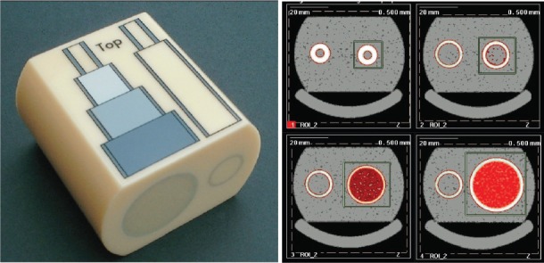
The choice of an appropriate imaging technique to quantify bone, muscle, or muscle adiposity needs to be guided by a thorough understanding of its competitive advantages over other modalities balanced by its limitations. This review details the technical machinery and methods behind peripheral quantitative computed tomography (pQCT), high-resolution (HR)-pQCT, and magnetic resonance imaging (MRI) that drive successful depiction of bone and muscle morphometry, densitometry, and structure. It discusses a number of image acquisition settings, the challenges associated with using one versus another, and compares the risk-benefits across the different modalities. Issues related to all modalities including partial volume artifact, beam hardening, calibration, and motion assessment are also detailed. The review further provides data and images to illustrate differences between methods to better guide the reader in selecting an imaging method strategically. Overall, investigators should be cautious of the impact of imaging parameters on image signal or contrast-to-noise-ratios, and the need to report these settings in future publications. The effect of motion should be assessed on images and a decision made to exclude prior to segmentation. A more standardized approach to imaging bone and muscle on pQCT and MRI could enhance comparability across studies and could improve the quality of meta-analyses.

摘要

选择合适的成像技术来量化骨骼、肌肉或肌肉脂肪含量,需要在全面了解其相对于其他模式的竞争优势及其局限性之间取得平衡的基础上进行指导。本综述详细介绍了外周定量计算机断层扫描(pQCT)、高分辨率(HR)-pQCT和磁共振成像(MRI)背后的技术机制和方法,这些技术可成功描绘骨骼和肌肉的形态测量、密度测量和结构。它讨论了一些图像采集设置、使用一种技术与另一种技术相关的挑战,并比较了不同模式的风险效益。还详细介绍了与所有模式相关的问题,包括部分容积伪影、束硬化、校准和运动评估。本综述进一步提供数据和图像,以说明不同方法之间的差异,从而更好地指导读者战略性地选择成像方法。总体而言,研究人员应谨慎对待成像参数对图像信号或对比度噪声比的影响,以及在未来出版物中报告这些设置的必要性。应在图像上评估运动的影响,并在分割前决定是否排除。采用更标准化的方法在pQCT和MRI上对骨骼和肌肉进行成像,可以提高不同研究之间的可比性,并改善荟萃分析的质量。

https://cdn.ncbi.nlm.nih.gov/pmc/blobs/06aa/5259568/bb7ab387a6b8/JMNI-16-265-g013.jpg
https://cdn.ncbi.nlm.nih.gov/pmc/blobs/06aa/5259568/fd28b6ca8490/JMNI-16-265-g001.jpg
https://cdn.ncbi.nlm.nih.gov/pmc/blobs/06aa/5259568/e5c217e3d84b/JMNI-16-265-g003.jpg
https://cdn.ncbi.nlm.nih.gov/pmc/blobs/06aa/5259568/1bfd2a4f913c/JMNI-16-265-g004.jpg
https://cdn.ncbi.nlm.nih.gov/pmc/blobs/06aa/5259568/27750a9eac13/JMNI-16-265-g006.jpg
https://cdn.ncbi.nlm.nih.gov/pmc/blobs/06aa/5259568/fb724a0c9a7f/JMNI-16-265-g007.jpg
https://cdn.ncbi.nlm.nih.gov/pmc/blobs/06aa/5259568/ee2159626193/JMNI-16-265-g008.jpg
https://cdn.ncbi.nlm.nih.gov/pmc/blobs/06aa/5259568/81557a2682a7/JMNI-16-265-g009.jpg
https://cdn.ncbi.nlm.nih.gov/pmc/blobs/06aa/5259568/ee3006e18ae3/JMNI-16-265-g010.jpg
https://cdn.ncbi.nlm.nih.gov/pmc/blobs/06aa/5259568/59ce4e08b05d/JMNI-16-265-g011.jpg
https://cdn.ncbi.nlm.nih.gov/pmc/blobs/06aa/5259568/2b88622bb40f/JMNI-16-265-g012.jpg
https://cdn.ncbi.nlm.nih.gov/pmc/blobs/06aa/5259568/bb7ab387a6b8/JMNI-16-265-g013.jpg

相似文献

1
A comparison of peripheral imaging technologies for bone and muscle quantification: a technical review of image acquisition.用于骨骼和肌肉定量分析的外周成像技术比较:图像采集技术综述
J Musculoskelet Neuronal Interact. 2016 Dec 14;16(4):265-282.
2
A new algorithm to improve assessment of cortical bone geometry in pQCT.一种用于改进外周定量计算机断层扫描中皮质骨几何结构评估的新算法。
Bone. 2015 Dec;81:721-730. doi: 10.1016/j.bone.2015.09.015. Epub 2015 Sep 30.
3
Human trabecular bone microarchitecture can be assessed independently of density with second generation HR-pQCT.利用第二代高分辨率外周定量计算机断层扫描(HR-pQCT),可以独立于骨密度评估人类小梁骨微结构。
Bone. 2015 Oct;79:213-21. doi: 10.1016/j.bone.2015.06.006. Epub 2015 Jun 14.
4
Automated quantification of three-dimensional subject motion to monitor image quality in high-resolution peripheral quantitative computed tomography.自动化三维受试者运动定量分析,以监测高分辨率外周定量 CT 中的图像质量。
Phys Med Biol. 2011 Oct 21;56(20):6523-43. doi: 10.1088/0031-9155/56/20/001. Epub 2011 Sep 21.
5
A Comparison of Peripheral Imaging Technologies for Bone and Muscle Quantification: a Mixed Methods Clinical Review.用于骨骼和肌肉定量的外周成像技术比较:一项混合方法的临床综述
Curr Osteoporos Rep. 2016 Dec;14(6):359-373. doi: 10.1007/s11914-016-0334-z.
6
A Comparison of Peripheral Imaging Technologies for Bone and Muscle Quantification: A Review of Segmentation Techniques.外周影像学技术在骨骼和肌肉定量方面的比较:分割技术综述。
J Clin Densitom. 2020 Jan-Mar;23(1):92-107. doi: 10.1016/j.jocd.2018.04.001. Epub 2018 Apr 17.
7
Accuracy of high-resolution peripheral quantitative computed tomography for measurement of bone quality.高分辨率外周定量计算机断层扫描测量骨质量的准确性
Med Eng Phys. 2007 Dec;29(10):1096-105. doi: 10.1016/j.medengphy.2006.11.002. Epub 2007 Jan 16.
8
Technical Note: Evaluation of a 160-mm/256-row CT scanner for whole-heart quantitative myocardial perfusion imaging.技术说明:用于全心定量心肌灌注成像的160毫米/256排CT扫描仪的评估
Med Phys. 2016 Aug;43(8):4821. doi: 10.1118/1.4957389.
9
Bone Quality-Beyond Bone Mineral Density.骨质量——超越骨密度
Semin Musculoskelet Radiol. 2016 Jul;20(3):269-278. doi: 10.1055/s-0036-1592365. Epub 2016 Oct 14.
10
Assessment of 3-month changes in bone microstructure under anti-TNFα therapy in patients with rheumatoid arthritis using high-resolution peripheral quantitative computed tomography (HR-pQCT).采用高分辨率外周定量计算机断层扫描(HR-pQCT)评估类风湿关节炎患者抗 TNFα 治疗 3 个月后的骨微观结构变化。
Arthritis Res Ther. 2017 Oct 4;19(1):222. doi: 10.1186/s13075-017-1430-x.

引用本文的文献

1
Smaller and thinner long bones in children and adolescents with cerebral palsy and other neuromotor impairments.患有脑瘫和其他神经运动障碍的儿童及青少年的长骨更小、更细。
Front Endocrinol (Lausanne). 2025 Jun 30;16:1620573. doi: 10.3389/fendo.2025.1620573. eCollection 2025.
2
Bone imaging modality precision and agreement between DXA, pQCT, and HR-pQCT.双能X线吸收法(DXA)、外周定量CT(pQCT)和高分辨率外周定量CT(HR-pQCT)之间的骨成像模态精度与一致性。
JBMR Plus. 2024 Dec 3;9(2):ziae158. doi: 10.1093/jbmrpl/ziae158. eCollection 2025 Feb.
3
Use of noninvasive imaging to identify causes of skeletal fragility in adults with diabetes: a review.

本文引用的文献

1
A Comparison of Peripheral Imaging Technologies for Bone and Muscle Quantification: a Mixed Methods Clinical Review.用于骨骼和肌肉定量的外周成像技术比较:一项混合方法的临床综述
Curr Osteoporos Rep. 2016 Dec;14(6):359-373. doi: 10.1007/s11914-016-0334-z.
2
Relationships between fatty infiltration in the thigh and calf in women with knee osteoarthritis.膝关节骨关节炎女性患者大腿与小腿脂肪浸润之间的关系。
Aging Clin Exp Res. 2017 Apr;29(2):291-299. doi: 10.1007/s40520-016-0556-z. Epub 2016 Mar 10.
3
Impact of incorrect tissue classification in Dixon-based MR-AC: fat-water tissue inversion.
利用非侵入性成像技术识别糖尿病成人骨骼脆弱的原因:综述
JBMR Plus. 2024 Jan 19;8(2):ziae003. doi: 10.1093/jbmrpl/ziae003. eCollection 2024 Feb.
4
Bone Quality in Competitive Athletes: A Systematic Review.竞技运动员的骨质量:系统评价。
J Musculoskelet Neuronal Interact. 2023 Dec 1;23(4):456-470.
5
What are the Effects of Exercise on Trabecular Microarchitecture in Older Adults? A Systematic Review and Meta-analysis of HR-pQCT Studies.运动对老年人小梁骨微观结构有何影响?HR-pQCT 研究的系统评价和荟萃分析。
Calcif Tissue Int. 2023 Oct;113(4):359-382. doi: 10.1007/s00223-023-01127-7. Epub 2023 Sep 19.
6
Associations Between Aldosterone-Renin-Ratio and Bone Parameters Derived from Peripheral Quantitative Computed Tomography and Impact Microindentation in Men.男性外周定量计算机断层扫描和冲击微压痕测定的醛固酮-肾素-比值与骨参数的相关性。
Calcif Tissue Int. 2023 Nov;113(5):496-510. doi: 10.1007/s00223-023-01131-x. Epub 2023 Sep 10.
7
Deep learning in sex estimation from a peripheral quantitative computed tomography scan of the fourth lumbar vertebra-a proof-of-concept study.基于第四腰椎外周定量 CT 扫描的深度学习性别估计:概念验证研究。
Forensic Sci Med Pathol. 2023 Dec;19(4):534-540. doi: 10.1007/s12024-023-00586-6. Epub 2023 Feb 11.
8
Multi-atlas segmentation and quantification of muscle, bone and subcutaneous adipose tissue in the lower leg using peripheral quantitative computed tomography.使用外周定量计算机断层扫描对小腿肌肉、骨骼和皮下脂肪组织进行多图谱分割与量化
Front Physiol. 2022 Oct 14;13:951368. doi: 10.3389/fphys.2022.951368. eCollection 2022.
9
HR-pQCT for the Evaluation of Muscle Quality and Intramuscular Fat Infiltration in Ageing Skeletal Muscle.用于评估衰老骨骼肌中肌肉质量和肌内脂肪浸润的高分辨率外周定量计算机断层扫描
J Pers Med. 2022 Jun 20;12(6):1016. doi: 10.3390/jpm12061016.
10
Validation of High-Resolution peripheral Quantitative Computed Tomography-Derived Achilles Tendon Properties Against Diagnostic Ultrasound.高频外周定量计算机断层扫描衍生跟腱特性的验证与诊断超声相对照。
J Musculoskelet Neuronal Interact. 2022 Jun 1;22(2):203-211.
基于狄克逊技术的磁共振图像自动分类中错误组织分类的影响:脂肪-水组织反转
EJNMMI Phys. 2014 Dec;1(1):101. doi: 10.1186/s40658-014-0101-0. Epub 2014 Dec 14.
4
Field inhomogeneity correction using partial differential phases in magnetic resonance imaging.
Phys Med Biol. 2015 May 21;60(10):4075-88. doi: 10.1088/0031-9155/60/10/4075. Epub 2015 Apr 30.
5
T2 relaxation times are increased in Skeletal muscle of DMD but not BMD patients.杜氏肌营养不良症(DMD)患者的骨骼肌中T2弛豫时间增加,但贝克型肌营养不良症(BMD)患者则不然。
Muscle Nerve. 2016 Jan;53(1):38-43. doi: 10.1002/mus.24679. Epub 2015 Nov 26.
6
Associations between pQCT-based fat and muscle area and density and DXA-based total and leg soft tissue mass in healthy women and men.基于外周定量计算机断层扫描的脂肪和肌肉面积、密度与基于双能X线吸收法的健康女性和男性全身及腿部软组织质量之间的关联。
J Musculoskelet Neuronal Interact. 2014 Dec;14(4):411-7.
7
A trimodality comparison of volumetric bone imaging technologies. Part I: Short-term precision and validity.三种容积骨成像技术的对比。第一部分:短期精密度和准确性。
J Clin Densitom. 2015 Jan-Mar;18(1):124-35. doi: 10.1016/j.jocd.2014.07.005. Epub 2014 Aug 13.
8
Variable flip angle three-dimensional fast spin-echo sequence combined with outer volume suppression for imaging trabecular bone structure of the proximal femur.可变翻转角三维快速自旋回波序列联合外周容积抑制用于股骨近端小梁骨结构成像
J Magn Reson Imaging. 2015 May;41(5):1300-10. doi: 10.1002/jmri.24673. Epub 2014 Jun 23.
9
Standardizing evaluation of pQCT image quality in the presence of subject movement: qualitative versus quantitative assessment.在存在受试者运动的情况下对 pQCT 图像质量进行标准化评估:定性与定量评估。
Calcif Tissue Int. 2014 Feb;94(2):202-11. doi: 10.1007/s00223-013-9803-x.
10
A new method of segmentation of compact-appearing, transitional and trabecular compartments and quantification of cortical porosity from high resolution peripheral quantitative computed tomographic images.一种从高分辨率外周定量 CT 图像中分割外观紧凑、过渡和小梁骨腔并量化皮质孔隙率的新方法。
Bone. 2013 May;54(1):8-20. doi: 10.1016/j.bone.2013.01.007. Epub 2013 Jan 17.