• 文献检索
  • 文档翻译
  • 深度研究
  • 学术资讯
  • Suppr Zotero 插件Zotero 插件
  • 邀请有礼
  • 套餐&价格
  • 历史记录
应用&插件
Suppr Zotero 插件Zotero 插件浏览器插件Mac 客户端Windows 客户端微信小程序
定价
高级版会员购买积分包购买API积分包
服务
文献检索文档翻译深度研究API 文档MCP 服务
关于我们
关于 Suppr公司介绍联系我们用户协议隐私条款
关注我们

Suppr 超能文献

核心技术专利:CN118964589B侵权必究
粤ICP备2023148730 号-1Suppr @ 2026

文献检索

告别复杂PubMed语法,用中文像聊天一样搜索,搜遍4000万医学文献。AI智能推荐,让科研检索更轻松。

立即免费搜索

文件翻译

保留排版,准确专业,支持PDF/Word/PPT等文件格式,支持 12+语言互译。

免费翻译文档

深度研究

AI帮你快速写综述,25分钟生成高质量综述,智能提取关键信息,辅助科研写作。

立即免费体验

在三个乳腺摄影系统中,对配对的原始数字图像与处理后的数字图像以及配对的屏-片图像与数字图像进行乳腺X线密度评估。

Mammographic density assessed on paired raw and processed digital images and on paired screen-film and digital images across three mammography systems.

作者信息

Burton Anya, Byrnes Graham, Stone Jennifer, Tamimi Rulla M, Heine John, Vachon Celine, Ozmen Vahit, Pereira Ana, Garmendia Maria Luisa, Scott Christopher, Hipwell John H, Dickens Caroline, Schüz Joachim, Aribal Mustafa Erkin, Bertrand Kimberly, Kwong Ava, Giles Graham G, Hopper John, Pérez Gómez Beatriz, Pollán Marina, Teo Soo-Hwang, Mariapun Shivaani, Taib Nur Aishah Mohd, Lajous Martín, Lopez-Riduara Ruy, Rice Megan, Romieu Isabelle, Flugelman Anath Arzee, Ursin Giske, Qureshi Samera, Ma Huiyan, Lee Eunjung, Sirous Reza, Sirous Mehri, Lee Jong Won, Kim Jisun, Salem Dorria, Kamal Rasha, Hartman Mikael, Miao Hui, Chia Kee-Seng, Nagata Chisato, Vinayak Sudhir, Ndumia Rose, van Gils Carla H, Wanders Johanna O P, Peplonska Beata, Bukowska Agnieszka, Allen Steve, Vinnicombe Sarah, Moss Sue, Chiarelli Anna M, Linton Linda, Maskarinec Gertraud, Yaffe Martin J, Boyd Norman F, Dos-Santos-Silva Isabel, McCormack Valerie A

机构信息

Section of Environment and Radiation, International Agency for Research on Cancer, 150 cours Albert Thomas, 69372, Lyon, Cedex 09, France.

Centre for Genetic Origins of Health and Disease, Curtin University and the University of Western Australia, Perth, Australia.

出版信息

Breast Cancer Res. 2016 Dec 19;18(1):130. doi: 10.1186/s13058-016-0787-0.

DOI:10.1186/s13058-016-0787-0
PMID:27993168
原文链接:https://pmc.ncbi.nlm.nih.gov/articles/PMC5168805/
Abstract

BACKGROUND

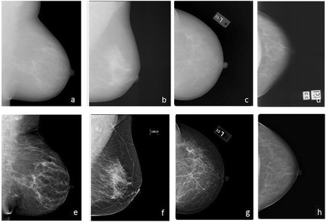
Inter-women and intra-women comparisons of mammographic density (MD) are needed in research, clinical and screening applications; however, MD measurements are influenced by mammography modality (screen film/digital) and digital image format (raw/processed). We aimed to examine differences in MD assessed on these image types.

METHODS

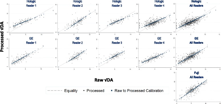
We obtained 1294 pairs of images saved in both raw and processed formats from Hologic and General Electric (GE) direct digital systems and a Fuji computed radiography (CR) system, and 128 screen-film and processed CR-digital pairs from consecutive screening rounds. Four readers performed Cumulus-based MD measurements (n = 3441), with each image pair read by the same reader. Multi-level models of square-root percent MD were fitted, with a random intercept for woman, to estimate processed-raw MD differences.

RESULTS

Breast area did not differ in processed images compared with that in raw images, but the percent MD was higher, due to a larger dense area (median 28.5 and 25.4 cm respectively, mean √dense area difference 0.44 cm (95% CI: 0.36, 0.52)). This difference in √dense area was significant for direct digital systems (Hologic 0.50 cm (95% CI: 0.39, 0.61), GE 0.56 cm (95% CI: 0.42, 0.69)) but not for Fuji CR (0.06 cm (95% CI: -0.10, 0.23)). Additionally, within each system, reader-specific differences varied in magnitude and direction (p < 0.001). Conversion equations revealed differences converged to zero with increasing dense area. MD differences between screen-film and processed digital on the subsequent screening round were consistent with expected time-related MD declines.

CONCLUSIONS

MD was slightly higher when measured on processed than on raw direct digital mammograms. Comparisons of MD on these image formats should ideally control for this non-constant and reader-specific difference.

摘要

背景

在研究、临床及筛查应用中,需要对女性之间以及女性个体内部的乳腺X线密度(MD)进行比较;然而,MD测量受乳腺X线摄影方式(屏-片/数字)及数字图像格式(原始/处理后)的影响。我们旨在研究这些图像类型上评估的MD差异。

方法

我们从Hologic和通用电气(GE)直接数字系统以及富士计算机X线摄影(CR)系统中获取了1294对以原始和处理后格式保存的图像,以及来自连续筛查轮次的128对屏-片和处理后的CR-数字图像。四位阅片者进行基于积云的MD测量(n = 3441),每对图像由同一位阅片者阅读。对平方根百分比MD拟合多级模型,对女性采用随机截距,以估计处理后-原始MD差异。

结果

与原始图像相比,处理后图像的乳房面积无差异,但MD百分比更高,这是由于致密面积更大(中位数分别为28.5和25.4平方厘米,致密面积平均差异的平方根为0.44厘米(95%可信区间:0.36,0.52))。这种致密面积平方根的差异在直接数字系统中显著(Hologic为0.50厘米(95%可信区间:0.39,0.61),GE为0.56厘米(95%可信区间:0.42,0.69)),但在富士CR系统中不显著(0.06厘米(95%可信区间:-0.10,0.23))。此外,在每个系统内,阅片者特异性差异在大小和方向上各不相同(p < 0.001)。转换方程显示,差异随着致密面积的增加而趋于零。后续筛查轮次中屏-片和处理后数字图像之间的MD差异与预期的与时间相关的MD下降一致。

结论

在处理后的直接数字乳腺X线摄影上测量的MD略高于原始图像。在这些图像格式上进行MD比较时,理想情况下应控制这种非恒定且阅片者特异性的差异。

https://cdn.ncbi.nlm.nih.gov/pmc/blobs/e21f/5168805/a1b334d59966/13058_2016_787_Fig2_HTML.jpg
https://cdn.ncbi.nlm.nih.gov/pmc/blobs/e21f/5168805/7bbe3cc8d0fb/13058_2016_787_Fig1_HTML.jpg
https://cdn.ncbi.nlm.nih.gov/pmc/blobs/e21f/5168805/a1b334d59966/13058_2016_787_Fig2_HTML.jpg
https://cdn.ncbi.nlm.nih.gov/pmc/blobs/e21f/5168805/7bbe3cc8d0fb/13058_2016_787_Fig1_HTML.jpg
https://cdn.ncbi.nlm.nih.gov/pmc/blobs/e21f/5168805/a1b334d59966/13058_2016_787_Fig2_HTML.jpg

相似文献

1
Mammographic density assessed on paired raw and processed digital images and on paired screen-film and digital images across three mammography systems.在三个乳腺摄影系统中,对配对的原始数字图像与处理后的数字图像以及配对的屏-片图像与数字图像进行乳腺X线密度评估。
Breast Cancer Res. 2016 Dec 19;18(1):130. doi: 10.1186/s13058-016-0787-0.
2
Impact of type of full-field digital image on mammographic density assessment and breast cancer risk estimation: a case-control study.全视野数字图像类型对乳腺密度评估和乳腺癌风险估计的影响:一项病例对照研究。
Breast Cancer Res. 2016 Sep 26;18(1):96. doi: 10.1186/s13058-016-0756-7.
3
Estimation of breast percent density in raw and processed full field digital mammography images via adaptive fuzzy c-means clustering and support vector machine segmentation.通过自适应模糊C均值聚类和支持向量机分割法估计原始及处理后的全视野数字化乳腺摄影图像中的乳腺密度百分比
Med Phys. 2012 Aug;39(8):4903-17. doi: 10.1118/1.4736530.
4
Comparison of percent density from raw and processed full-field digital mammography data.原始和处理后的全场数字化乳腺摄影数据的密度百分比比较。
Breast Cancer Res. 2013 Jan 4;15(1):R1. doi: 10.1186/bcr3372.
5
Case-control study of mammographic density and breast cancer risk using processed digital mammograms.利用处理后的数字化乳腺X线摄影图像进行乳腺密度与乳腺癌风险的病例对照研究。
Breast Cancer Res. 2016 May 21;18(1):53. doi: 10.1186/s13058-016-0715-3.
6
Reproductive Factors and Mammographic Density: Associations Among 24,840 Women and Comparison of Studies Using Digitized Film-Screen Mammography and Full-Field Digital Mammography.生殖因素与乳腺 X 线摄影密度:24840 名女性的相关性研究及数字化屏片乳腺摄影与全数字化乳腺摄影研究的比较
Am J Epidemiol. 2019 Jun 1;188(6):1144-1154. doi: 10.1093/aje/kwz033.
7
Evaluation of LIBRA Software for Fully Automated Mammographic Density Assessment in Breast Cancer Risk Prediction.评估 LIBRA 软件在乳腺癌风险预测中全自动乳腺密度评估的应用。
Radiology. 2020 Jul;296(1):24-31. doi: 10.1148/radiol.2020192509. Epub 2020 May 12.
8
Area and volumetric density estimation in processed full-field digital mammograms for risk assessment of breast cancer.用于乳腺癌风险评估的处理后全场数字化乳腺X线摄影中的面积和体积密度估计。
PLoS One. 2014 Oct 20;9(10):e110690. doi: 10.1371/journal.pone.0110690. eCollection 2014.
9
Cancer cases from ACRIN digital mammographic imaging screening trial: radiologist analysis with use of a logistic regression model.来自ACRIN数字化乳腺钼靶成像筛查试验的癌症病例:使用逻辑回归模型的放射科医生分析
Radiology. 2009 Aug;252(2):348-57. doi: 10.1148/radiol.2522081457.
10
Breast parenchymal patterns in processed versus raw digital mammograms: A large population study toward assessing differences in quantitative measures across image representations.处理后的数字乳腺钼靶图像与原始数字乳腺钼靶图像中的乳腺实质模式:一项针对评估不同图像表现形式下定量测量差异的大规模人群研究。
Med Phys. 2016 Nov;43(11):5862. doi: 10.1118/1.4963810.

引用本文的文献

1
Mammographic density and breast cancer risk among Black American women.美国黑人女性的乳腺X线密度与乳腺癌风险
Int J Cancer. 2025 Mar 15;156(6):1173-1180. doi: 10.1002/ijc.35223. Epub 2024 Oct 19.
2
Association and Prediction Utilizing Craniocaudal and Mediolateral Oblique View Digital Mammography and Long-Term Breast Cancer Risk.利用头尾位和内外斜位数字乳腺摄影术与长期乳腺癌风险的关联和预测。
Cancer Prev Res (Phila). 2023 Sep 1;16(9):531-537. doi: 10.1158/1940-6207.CAPR-22-0499.
3
The association of age at menarche and adult height with mammographic density in the International Consortium of Mammographic Density.

本文引用的文献

1
Impact of type of full-field digital image on mammographic density assessment and breast cancer risk estimation: a case-control study.全视野数字图像类型对乳腺密度评估和乳腺癌风险估计的影响:一项病例对照研究。
Breast Cancer Res. 2016 Sep 26;18(1):96. doi: 10.1186/s13058-016-0756-7.
2
International Consortium on Mammographic Density: Methodology and population diversity captured across 22 countries.国际乳腺密度联盟:22个国家的方法学与人群多样性
Cancer Epidemiol. 2016 Feb;40:141-51. doi: 10.1016/j.canep.2015.11.015. Epub 2015 Dec 24.
3
Mammographic density defined by higher than conventional brightness threshold better predicts breast cancer risk for full-field digital mammograms.
国际乳腺密度研究联合会中月经初潮年龄和成人身高与乳腺密度的相关性研究。
Breast Cancer Res. 2022 Jul 14;24(1):49. doi: 10.1186/s13058-022-01545-9.
4
Deep learning identification of stiffness markers in breast cancer.深度学习识别乳腺癌的硬度标志物。
Biomaterials. 2022 Jun;285:121540. doi: 10.1016/j.biomaterials.2022.121540. Epub 2022 Apr 27.
5
Mammographic texture features associated with contralateral breast cancer in the WECARE Study.WECARE研究中与对侧乳腺癌相关的乳腺X线纹理特征。
NPJ Breast Cancer. 2021 Nov 29;7(1):146. doi: 10.1038/s41523-021-00354-1.
6
A Deep Learning Approach to Re-create Raw Full-Field Digital Mammograms for Breast Density and Texture Analysis.一种用于重新创建原始全场数字化乳腺X线摄影图像以进行乳腺密度和纹理分析的深度学习方法。
Radiol Artif Intell. 2021 Apr 14;3(4):e200097. doi: 10.1148/ryai.2021200097. eCollection 2021 Jul.
7
The distribution and determinants of mammographic density measures in Western Australian aboriginal women.西澳大利亚原住民妇女的乳腺 X 光密度测量分布及其决定因素。
Breast Cancer Res. 2019 Feb 28;21(1):33. doi: 10.1186/s13058-019-1113-4.
8
A comprehensive tool for measuring mammographic density changes over time.一种全面的工具,用于测量随时间变化的乳腺密度变化。
Breast Cancer Res Treat. 2018 Jun;169(2):371-379. doi: 10.1007/s10549-018-4690-5. Epub 2018 Feb 1.
9
Automated mammographic breast density estimation using a fully convolutional network.使用全卷积网络进行自动乳腺钼靶密度估计。
Med Phys. 2018 Mar;45(3):1178-1190. doi: 10.1002/mp.12763. Epub 2018 Feb 19.
10
Mammographic density and ageing: A collaborative pooled analysis of cross-sectional data from 22 countries worldwide.乳腺X线密度与衰老:来自全球22个国家横断面数据的联合汇总分析。
PLoS Med. 2017 Jun 30;14(6):e1002335. doi: 10.1371/journal.pmed.1002335. eCollection 2017 Jun.
通过高于传统亮度阈值定义的乳腺X线摄影密度能更好地预测全视野数字化乳腺X线摄影的乳腺癌风险。
Breast Cancer Res. 2015 Nov 18;17:142. doi: 10.1186/s13058-015-0654-4.
4
Preliminary evaluation of the publicly available Laboratory for Breast Radiodensity Assessment (LIBRA) software tool: comparison of fully automated area and volumetric density measures in a case-control study with digital mammography.乳腺放射密度评估实验室(LIBRA)软件工具的初步评估:在一项数字乳腺摄影病例对照研究中全自动面积和体积密度测量的比较。
Breast Cancer Res. 2015 Aug 25;17:117. doi: 10.1186/s13058-015-0626-8.
5
Mammographic density measurements are not affected by mammography system.乳房X线密度测量不受乳房X线摄影系统的影响。
J Med Imaging (Bellingham). 2015 Jan;2(1):015501. doi: 10.1117/1.JMI.2.1.015501. Epub 2015 Mar 4.
6
Ethnic differences in mammographic densities: an Asian cross-sectional study.乳房X线密度的种族差异:一项亚洲横断面研究。
PLoS One. 2015 Feb 6;10(2):e0117568. doi: 10.1371/journal.pone.0117568. eCollection 2015.
7
Mammographic breast density refines Tyrer-Cuzick estimates of breast cancer risk in high-risk women: findings from the placebo arm of the International Breast Cancer Intervention Study I.乳腺钼靶密度可优化高危女性乳腺癌风险的 Tyrer-Cuzick 评估:国际乳腺癌干预研究 I 安慰剂组的研究结果
Breast Cancer Res. 2014 Oct 8;16(5):451. doi: 10.1186/s13058-014-0451-5.
8
Digital mammographic density and breast cancer risk: a case-control study of six alternative density assessment methods.数字化乳腺密度与乳腺癌风险:六种替代密度评估方法的病例对照研究
Breast Cancer Res. 2014 Sep 20;16(5):439. doi: 10.1186/s13058-014-0439-1.
9
Cancer incidence and mortality worldwide: sources, methods and major patterns in GLOBOCAN 2012.全球癌症发病与死亡:GLOBOCAN 2012 数据源、方法与主要模式。
Int J Cancer. 2015 Mar 1;136(5):E359-86. doi: 10.1002/ijc.29210. Epub 2014 Oct 9.
10
Successful first round results of a Turkish breast cancer screening program with mammography in Bahcesehir, Istanbul.在伊斯坦布尔巴赫切希尔开展的土耳其乳腺癌钼靶筛查项目首轮取得成功。
Asian Pac J Cancer Prev. 2014;15(4):1693-7. doi: 10.7314/apjcp.2014.15.4.1693.