Hejazi Fatemeh, Mirzadeh Hamid
Department of Polymer Engineering and Color Technology, Amirkabir University of Technology (Tehran Polytechnic), 424 Hafez Avenue, 1591634311, Tehran, Iran.
Prog Biomater. 2016 Dec;5(3-4):199-211. doi: 10.1007/s40204-016-0058-2. Epub 2016 Nov 18.
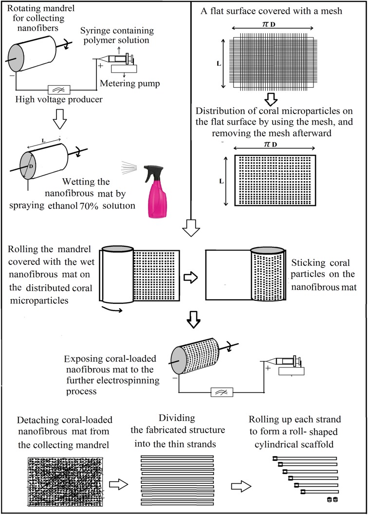
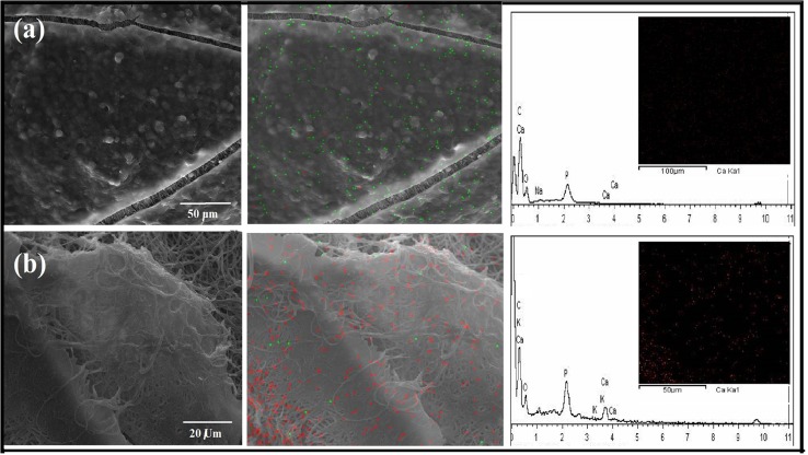
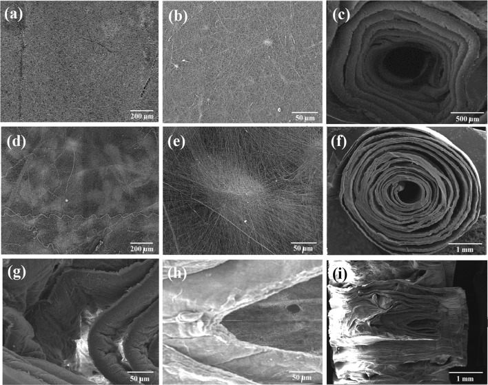
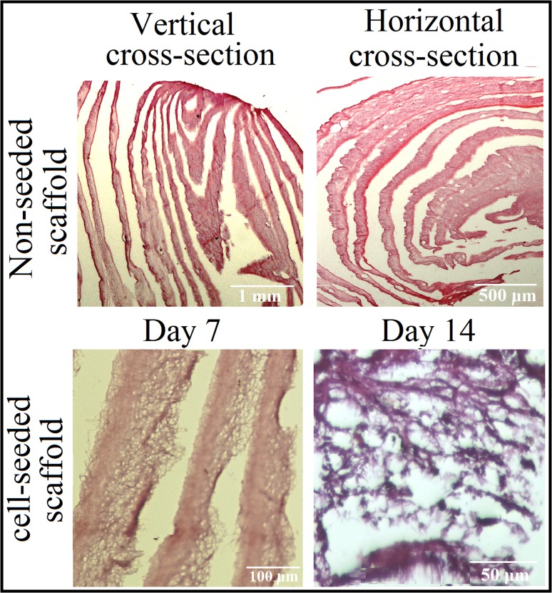
In this work, an innovative and easy method for the fabrication of 3D scaffold from 2D electrospun structures is introduced. For this aim, coral microparticles were fixed inside the nanofibrous PCL/Gelatin mat and the obtained structure was post assembled into a cylindrical design. Scaffold fabrication procedure is described in detail and morphological properties, physical and mechanical characteristics and in vitro assessments of the prepared scaffold are reported. Presences of coral microparticles in the structure led to the formation of empty spaces (3D pores) between nanofibrous layers which in turn prevent the compact accumulation of nanofibers. Post-assembly of the obtained nanofibrous coral-loaded structures makes it possible to prepare a scaffold with any desired dimension (diameter and height). Existence of coral particles within the nanofibrous mats resulted in distant placement of layers toward each other in the assembling step, which in turn create vacancy in the structure for cellular migration and fluid and nutrients exchange of the scaffold with the surrounding environment. Cell morphology within the scaffolds is investigated and cytotoxicity and cytocompatibility of the structure is evaluated using Alamar blue assay. Enhancement in mineralization of the seeded cells within the prepared coral-loaded scaffolds is demonstrated by the use of SEM-EDX. Performed compression mechanical test revealed excellent modulus and stiffness values for the cylindrical samples which are comparable to those of natural bone tissue.
在这项工作中,介绍了一种由二维电纺结构制造三维支架的创新且简便的方法。为此,将珊瑚微粒固定在纳米纤维聚己内酯/明胶垫内,并将所得结构后组装成圆柱形设计。详细描述了支架制造过程,并报告了所制备支架的形态特性、物理和机械特性以及体外评估结果。结构中珊瑚微粒的存在导致纳米纤维层之间形成空隙(三维孔隙),这反过来又阻止了纳米纤维的紧密堆积。所得负载珊瑚的纳米纤维结构的后组装使得制备具有任何所需尺寸(直径和高度)的支架成为可能。纳米纤维垫内珊瑚颗粒的存在导致在组装步骤中层彼此相距较远,这反过来又在结构中产生空位,用于细胞迁移以及支架与周围环境之间的流体和营养物质交换。研究了支架内的细胞形态,并使用阿拉玛蓝测定法评估了该结构的细胞毒性和细胞相容性。通过扫描电子显微镜-能谱分析(SEM-EDX)证明了制备的负载珊瑚支架内接种细胞矿化的增强。进行的压缩力学测试显示圆柱形样品具有优异的模量和刚度值,与天然骨组织相当。