From the *Department of Anesthesiology, Intensive Care and Pain Medicine, Tan Tock Seng Hospital, Singapore; †Department of Anesthesiology and Intensive Care Medicine, Aarhus University Hospital, Aarhus, Denmark; and ‡Department of Anesthesiology and Intensive Care Medicine, Zealand University Hospital, University of Copenhagen, Copenhagen, Denmark.
Reg Anesth Pain Med. 2017 Mar/Apr;42(2):241-245. doi: 10.1097/AAP.0000000000000539.
The precise location of the adductor canal remains controversial among anesthesiologists. In numerous studies of the analgesic effect of the so-called adductor canal block for total knee arthroplasty, the needle insertion point has been the midpoint of the thigh, determined as the midpoint between the anterior superior iliac spine and base of patella. "Adductor canal block" may be a misnomer for an approach that is actually an injection into the femoral triangle, a "femoral triangle block." This block probably has a different analgesic effect compared with an injection into the adductor canal. We sought to determine the exact location of the adductor canal using ultrasound and relate it to the midpoint of the thigh.
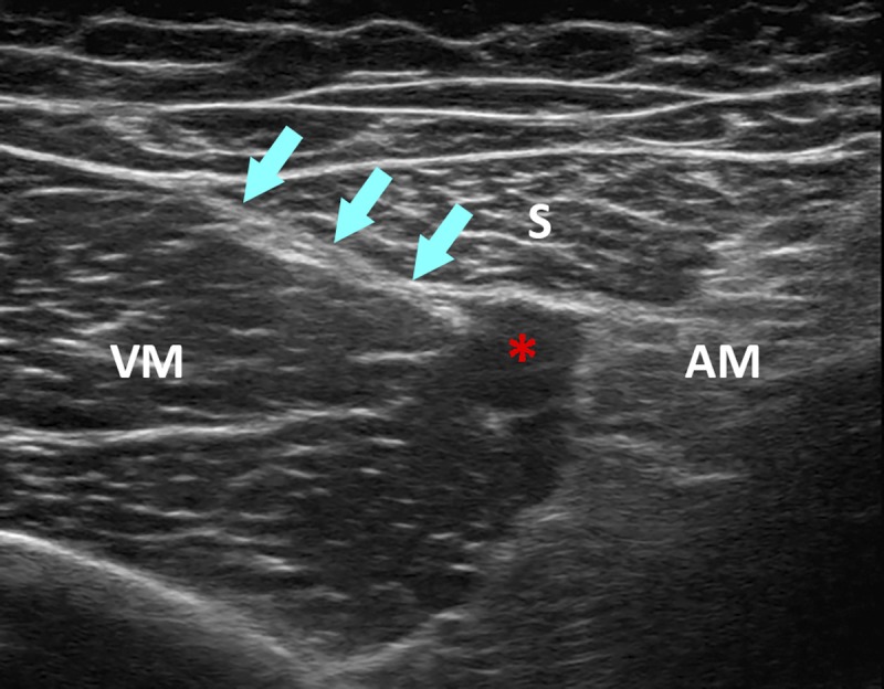
Twenty-two volunteers were examined using ultrasound. The proximal end of the adductor canal was identified where the medial border of the sartorius muscle intersects the medial border of the adductor longus muscle. The distal end of the adductor canal is the adductor hiatus, which was also visualized ultrasonographically.
The mean distance from the anterior superior iliac spine to the midpoint of the thigh was 22.9 cm (range, 20.3-24.9 cm). The mean distance from the anterior superior iliac spine to the proximal end of the adductor canal was 27.4 cm (range, 24.0-31.4 cm). Consequently, the mean distance from the midpoint of the thigh to the proximal end of the adductor canal was 4.6 cm (range, 2.3-7.0 cm).
In all volunteers, the midpoint of the thigh was proximal to the beginning of the adductor canal, suggesting that an injection performed at this level is in fact a femoral triangle block.
在麻醉医师中,收肌管的确切位置仍存在争议。在众多研究中,所谓的收肌管阻滞在全膝关节置换术中的镇痛效果,其进针点为大腿中部,即髂前上棘与髌骨基底的中点。“收肌管阻滞”可能是一种误称,因为这种方法实际上是注射到股三角,即“股三角阻滞”。与注射到收肌管相比,这种阻滞可能具有不同的镇痛效果。我们试图通过超声确定收肌管的确切位置,并将其与大腿中部相关联。
对 22 名志愿者进行超声检查。收肌管的近端在缝匠肌的内侧缘与收肌长肌的内侧缘相交处确定。收肌管的远端是收肌裂孔,也可以通过超声显示。
从髂前上棘到大腿中部的平均距离为 22.9cm(范围,20.3-24.9cm)。从髂前上棘到收肌管近端的平均距离为 27.4cm(范围,24.0-31.4cm)。因此,从大腿中部到收肌管近端的平均距离为 4.6cm(范围,2.3-7.0cm)。
在所有志愿者中,大腿中部位于收肌管起始部的近端,这表明在此水平进行的注射实际上是股三角阻滞。