• 文献检索
  • 文档翻译
  • 深度研究
  • 学术资讯
  • Suppr Zotero 插件Zotero 插件
  • 邀请有礼
  • 套餐&价格
  • 历史记录
应用&插件
Suppr Zotero 插件Zotero 插件浏览器插件Mac 客户端Windows 客户端微信小程序
定价
高级版会员购买积分包购买API积分包
服务
文献检索文档翻译深度研究API 文档MCP 服务
关于我们
关于 Suppr公司介绍联系我们用户协议隐私条款
关注我们

Suppr 超能文献

核心技术专利:CN118964589B侵权必究
粤ICP备2023148730 号-1Suppr @ 2026

文献检索

告别复杂PubMed语法,用中文像聊天一样搜索,搜遍4000万医学文献。AI智能推荐,让科研检索更轻松。

立即免费搜索

文件翻译

保留排版,准确专业,支持PDF/Word/PPT等文件格式,支持 12+语言互译。

免费翻译文档

深度研究

AI帮你快速写综述,25分钟生成高质量综述,智能提取关键信息,辅助科研写作。

立即免费体验

相似文献

1
DISCORDANCE BETWEEN BLUE-LIGHT AUTOFLUORESCENCE AND NEAR-INFRARED AUTOFLUORESCENCE IN AGE-RELATED MACULAR DEGENERATION.年龄相关性黄斑变性中蓝光自发荧光与近红外自发荧光之间的不一致性
Retina. 2016 Dec;36 Suppl 1(Suppl 1):S137-S146. doi: 10.1097/IAE.0000000000001254.
2
Fundus autofluorescence characteristics of nascent geographic atrophy in age-related macular degeneration.年龄相关性黄斑变性中新生地图样萎缩的眼底自发荧光特征
Invest Ophthalmol Vis Sci. 2015 Feb 12;56(3):1546-52. doi: 10.1167/iovs.14-16211.
3
Green emission fluorophores in eyes with atrophic age-related macular degeneration: a colour fundus autofluorescence pilot study.萎缩性年龄相关性黄斑变性眼的绿色发射荧光团:一项彩色眼底自发荧光初步研究。
Br J Ophthalmol. 2018 Jun;102(6):827-832. doi: 10.1136/bjophthalmol-2017-310881. Epub 2017 Sep 25.
4
CLINICOPATHOLOGIC CORRELATION OF GEOGRAPHIC ATROPHY SECONDARY TO AGE-RELATED MACULAR DEGENERATION.年龄相关性黄斑变性所致地图状萎缩的临床病理相关性。
Retina. 2019 Apr;39(4):802-816. doi: 10.1097/IAE.0000000000002461.
5
Optical coherence tomography-defined changes preceding the development of drusen-associated atrophy in age-related macular degeneration.光学相干断层扫描定义的在年龄相关性黄斑变性相关的 drusen 相关萎缩发展之前的变化。
Ophthalmology. 2014 Dec;121(12):2415-22. doi: 10.1016/j.ophtha.2014.06.034. Epub 2014 Aug 8.
6
HYPERSPECTRAL AUTOFLUORESCENCE IMAGING OF DRUSEN AND RETINAL PIGMENT EPITHELIUM IN DONOR EYES WITH AGE-RELATED MACULAR DEGENERATION.年龄相关性黄斑变性供体眼玻璃膜疣和视网膜色素上皮的高光谱自发荧光成像
Retina. 2016 Dec;36 Suppl 1(Suppl 1):S127-S136. doi: 10.1097/IAE.0000000000001325.
7
Autofluorescence characteristics of early, atrophic, and high-risk fellow eyes in age-related macular degeneration.年龄相关性黄斑变性早期、萎缩性和高危对侧眼的自发荧光特征
Invest Ophthalmol Vis Sci. 2006 Dec;47(12):5495-504. doi: 10.1167/iovs.05-1318.
8
Evaluation of intraretinal migration of retinal pigment epithelial cells in age-related macular degeneration using polarimetric imaging.应用偏光成像评估年龄相关性黄斑变性的视网膜色素上皮细胞的视网膜内迁移。
Sci Rep. 2017 Jun 9;7(1):3150. doi: 10.1038/s41598-017-03529-8.
9
In-vivo mapping of drusen by fundus autofluorescence and spectral-domain optical coherence tomography imaging.通过眼底自发荧光和光谱域光学相干断层扫描成像对玻璃膜疣进行体内成像。
Graefes Arch Clin Exp Ophthalmol. 2016 Jan;254(1):59-67. doi: 10.1007/s00417-015-3012-4. Epub 2015 Apr 24.
10
Microperimetric correlations of autofluorescence and optical coherence tomography imaging in dry age-related macular degeneration.干性年龄相关性黄斑变性的自发荧光与光相干断层扫描成像的微视野相关性。
Am J Ophthalmol. 2012 Jun;153(6):1110-5. doi: 10.1016/j.ajo.2011.11.002. Epub 2012 Feb 8.

引用本文的文献

1
Short-Wavelength and Infrared Autofluorescence Imaging in Pachychoroid Neovasculopathy.肥厚性脉络膜新生血管病变中的短波和红外自发荧光成像
Vision (Basel). 2025 Apr 21;9(2):38. doi: 10.3390/vision9020038.
2
Fundus Autofluorescence Variation in Geographic Atrophy of Age-Related Macular Degeneration: A Clinicopathologic Correlation.年龄相关性黄斑变性地图样萎缩中的眼底自发荧光变化:临床病理相关性研究
Invest Ophthalmol Vis Sci. 2025 Jan 2;66(1):49. doi: 10.1167/iovs.66.1.49.
3
Are the Hypo-Reflective Clumps Associated With Age-Related Macular Degeneration in Adaptive Optics Ophthalmoscopy Autofluorescent?自适应光学眼底镜检查中与年龄相关性黄斑变性相关的低反射团块是否为自发荧光?
Invest Ophthalmol Vis Sci. 2024 Aug 1;65(10):28. doi: 10.1167/iovs.65.10.28.
4
Adaptive Optics Retinal Imaging in RDH12-Associated Early Onset Severe Retinal Dystrophy.RDH12相关早发性严重视网膜营养不良的自适应光学视网膜成像
Invest Ophthalmol Vis Sci. 2024 Mar 5;65(3):9. doi: 10.1167/iovs.65.3.9.
5
SynthEye: Investigating the Impact of Synthetic Data on Artificial Intelligence-assisted Gene Diagnosis of Inherited Retinal Disease.SynthEye:研究合成数据对遗传性视网膜疾病人工智能辅助基因诊断的影响。
Ophthalmol Sci. 2022 Nov 22;3(2):100258. doi: 10.1016/j.xops.2022.100258. eCollection 2023 Jun.
6
The Fovea-Protective Impact of Double-Layer Sign in Eyes With Foveal-Sparing Geographic Atrophy and Age-Related Macular Degeneration.双层信号对具有中心凹 spared 型地理萎缩和年龄相关性黄斑变性的眼睛的中心凹保护作用。
Invest Ophthalmol Vis Sci. 2022 Oct 3;63(11):4. doi: 10.1167/iovs.63.11.4.
7
Long Term Time-Lapse Imaging of Geographic Atrophy: A Pilot Study.地图状萎缩的长期延时成像:一项初步研究。
Front Med (Lausanne). 2022 Jun 22;9:868163. doi: 10.3389/fmed.2022.868163. eCollection 2022.
8
Structural evaluation in inherited retinal diseases.遗传性视网膜疾病的结构评估。
Br J Ophthalmol. 2021 Dec;105(12):1623-1631. doi: 10.1136/bjophthalmol-2021-319228. Epub 2021 May 12.
9
Microstructure of the retinal pigment epithelium near-infrared autofluorescence in healthy young eyes and in patients with AMD.健康年轻眼睛和 AMD 患者的视网膜色素上皮层近红外自发荧光的微观结构。
Sci Rep. 2020 Jun 12;10(1):9561. doi: 10.1038/s41598-020-66581-x.
10
Calcified nodules in retinal drusen are associated with disease progression in age-related macular degeneration.视网膜玻璃膜疣中的钙化结节与年龄相关性黄斑变性的疾病进展有关。
Sci Transl Med. 2018 Nov 7;10(466). doi: 10.1126/scitranslmed.aat4544.

本文引用的文献

1
Near-Infrared Autofluorescence Imaging in Geographic Atrophy Using Spectralis Single and Combined Wavelength Modes.
Asia Pac J Ophthalmol (Phila). 2015 Nov-Dec;4(6):334-8. doi: 10.1097/APO.0000000000000142.
2
The Onion Sign in Neovascular Age-Related Macular Degeneration Represents Cholesterol Crystals.新生血管性年龄相关性黄斑变性中的洋葱征代表胆固醇结晶。
Ophthalmology. 2015 Nov;122(11):2316-26. doi: 10.1016/j.ophtha.2015.07.008. Epub 2015 Aug 19.
3
Flecks in Recessive Stargardt Disease: Short-Wavelength Autofluorescence, Near-Infrared Autofluorescence, and Optical Coherence Tomography.隐性Stargardt病中的斑点:短波自发荧光、近红外自发荧光和光学相干断层扫描
Invest Ophthalmol Vis Sci. 2015 Jul;56(8):5029-39. doi: 10.1167/iovs.15-16763.
4
Subretinal Hyperreflective Material in the Comparison of Age-Related Macular Degeneration Treatments Trials.年龄相关性黄斑变性治疗试验比较中的视网膜下高反射物质
Ophthalmology. 2015 Sep;122(9):1846-53.e5. doi: 10.1016/j.ophtha.2015.05.042. Epub 2015 Jul 2.
5
Autofluorescence imaging with near-infrared excitation:normalization by reflectance to reduce signal from choroidal fluorophores.近红外激发的自体荧光成像:通过反射率进行归一化以减少脉络膜荧光团的信号。
Invest Ophthalmol Vis Sci. 2015 May;56(5):3393-406. doi: 10.1167/iovs.15-16726.
6
REFRACTILE DRUSEN: Clinical Imaging and Candidate Histology.玻璃膜疣:临床成像与候选组织学
Retina. 2015 May;35(5):859-65. doi: 10.1097/IAE.0000000000000503.
7
Fundus autofluorescence imaging: Fundamentals and clinical relevance.眼底自发荧光成像:基础与临床意义。
Saudi J Ophthalmol. 2014 Apr;28(2):111-6. doi: 10.1016/j.sjopt.2014.03.008. Epub 2014 Mar 24.
8
Subretinal hyperreflective exudation associated with neovascular age-related macular degeneration.与新生血管性年龄相关性黄斑变性相关的视网膜下高反射性渗出物。
Retina. 2014 Jul;34(7):1281-8. doi: 10.1097/IAE.0000000000000166.
9
Increased fundus autofluorescence related to outer retinal disruption.与外层视网膜破坏相关的眼底自发荧光增强。
JAMA Ophthalmol. 2013 Dec;131(12):1645-9. doi: 10.1001/jamaophthalmol.2013.5030.
10
Quantitative fundus autofluorescence in healthy eyes.健康眼的定量眼底自发荧光。
Invest Ophthalmol Vis Sci. 2013 Aug 21;54(8):5684-93. doi: 10.1167/iovs.13-12445.

年龄相关性黄斑变性中蓝光自发荧光与近红外自发荧光之间的不一致性

DISCORDANCE BETWEEN BLUE-LIGHT AUTOFLUORESCENCE AND NEAR-INFRARED AUTOFLUORESCENCE IN AGE-RELATED MACULAR DEGENERATION.

作者信息

Heiferman Michael J, Fawzi Amani A

机构信息

Department of Ophthalmology, Northwestern University Feinberg School of Medicine, Chicago, Illinois.

出版信息

Retina. 2016 Dec;36 Suppl 1(Suppl 1):S137-S146. doi: 10.1097/IAE.0000000000001254.

DOI:10.1097/IAE.0000000000001254
PMID:28005672
原文链接:https://pmc.ncbi.nlm.nih.gov/articles/PMC5193230/
Abstract

PURPOSE

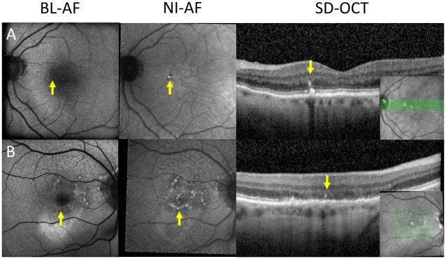
To identify the origin and significance of discordance between blue-light autofluorescence (BL-AF; 488 nm) and near-infrared autofluorescence (NI-AF; 787 nm) in patients with age-related macular degeneration (AMD).

METHODS

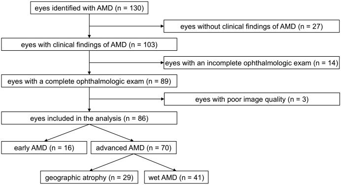
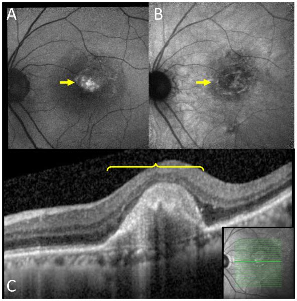
A total of 86 eyes of 59 patients with a diagnosis of AMD were included in this cross-sectional study conducted between March 9, 2015 and May 1, 2015. A masked observer examined the BL-AF, NI-AF, and spectral-domain optical coherence tomography images. Areas with discordance of autofluorescence patterns between NI-AF and BL-AF images were correlated with structural findings at the corresponding location in optical coherence tomography scans.

RESULTS

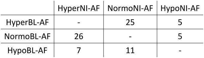
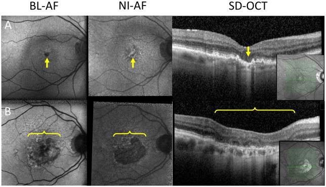
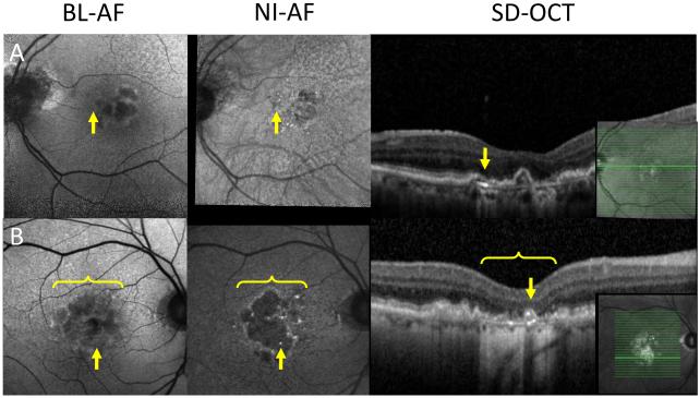
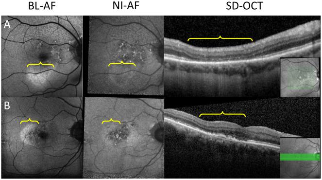
Seventy-nine eyes had discordance between BL-AF and NI-AF. The most common optical coherence tomography finding accounting for these discrepancies was pigment migration accounting for 35 lesions in 21 eyes. The most clinically relevant finding was geographic atrophy missed on BL-AF in 7 eyes.

CONCLUSION

Our findings indicate that variations in the distribution of lipofuscin, melanin and melanolipofuscin account for the majority of discordance between BL-AF and NI-AF. Given our finding of missed geographic atrophy lesions on BL-AF in 24% of eyes with geographic atrophy (7/29 eyes), clinicians should consider multimodal imaging, including NI-AF and optical coherence tomography, especially in clinical trials of geographic atrophy.

摘要

目的

确定年龄相关性黄斑变性(AMD)患者蓝光自发荧光(BL-AF;488nm)与近红外自发荧光(NI-AF;787nm)不一致的来源及意义。

方法

本横断面研究纳入了2015年3月9日至2015年5月1日期间诊断为AMD的59例患者的86只眼。一名经过遮蔽的观察者检查了BL-AF、NI-AF和光谱域光学相干断层扫描图像。NI-AF和BL-AF图像之间自发荧光模式不一致的区域与光学相干断层扫描相应位置的结构发现相关联。

结果

79只眼的BL-AF和NI-AF之间存在不一致。导致这些差异的最常见光学相干断层扫描发现是色素迁移,21只眼中有35个病灶。最具临床相关性的发现是7只眼中BL-AF遗漏了地图样萎缩。

结论

我们的研究结果表明,脂褐素、黑色素和黑素脂褐素分布的变化是BL-AF和NI-AF之间大部分不一致的原因。鉴于我们发现在24%的地图样萎缩眼中(7/29只眼)BL-AF遗漏了地图样萎缩病灶,临床医生应考虑多模态成像,包括NI-AF和光学相干断层扫描,尤其是在地图样萎缩的临床试验中。

https://cdn.ncbi.nlm.nih.gov/pmc/blobs/8897/5193230/931bdefbca98/nihms-803257-f0007.jpg
https://cdn.ncbi.nlm.nih.gov/pmc/blobs/8897/5193230/d3195370d561/nihms-803257-f0001.jpg
https://cdn.ncbi.nlm.nih.gov/pmc/blobs/8897/5193230/43febdc05a32/nihms-803257-f0002.jpg
https://cdn.ncbi.nlm.nih.gov/pmc/blobs/8897/5193230/eeff56b43d7c/nihms-803257-f0003.jpg
https://cdn.ncbi.nlm.nih.gov/pmc/blobs/8897/5193230/07f080c4cd34/nihms-803257-f0004.jpg
https://cdn.ncbi.nlm.nih.gov/pmc/blobs/8897/5193230/28c996802a84/nihms-803257-f0005.jpg
https://cdn.ncbi.nlm.nih.gov/pmc/blobs/8897/5193230/2a49f2586300/nihms-803257-f0006.jpg
https://cdn.ncbi.nlm.nih.gov/pmc/blobs/8897/5193230/931bdefbca98/nihms-803257-f0007.jpg