• 文献检索
  • 文档翻译
  • 深度研究
  • 学术资讯
  • Suppr Zotero 插件Zotero 插件
  • 邀请有礼
  • 套餐&价格
  • 历史记录
应用&插件
Suppr Zotero 插件Zotero 插件浏览器插件Mac 客户端Windows 客户端微信小程序
定价
高级版会员购买积分包购买API积分包
服务
文献检索文档翻译深度研究API 文档MCP 服务
关于我们
关于 Suppr公司介绍联系我们用户协议隐私条款
关注我们

Suppr 超能文献

核心技术专利:CN118964589B侵权必究
粤ICP备2023148730 号-1Suppr @ 2026

文献检索

告别复杂PubMed语法,用中文像聊天一样搜索,搜遍4000万医学文献。AI智能推荐,让科研检索更轻松。

立即免费搜索

文件翻译

保留排版,准确专业,支持PDF/Word/PPT等文件格式,支持 12+语言互译。

免费翻译文档

深度研究

AI帮你快速写综述,25分钟生成高质量综述,智能提取关键信息,辅助科研写作。

立即免费体验

遗传性视网膜疾病的结构评估。

Structural evaluation in inherited retinal diseases.

机构信息

Moorfields Eye Hospital City Road Campus, London, UK.

UCL Institute of Ophthalmology, University College London, London, UK.

出版信息

Br J Ophthalmol. 2021 Dec;105(12):1623-1631. doi: 10.1136/bjophthalmol-2021-319228. Epub 2021 May 12.

DOI:10.1136/bjophthalmol-2021-319228
PMID:33980508
原文链接:https://pmc.ncbi.nlm.nih.gov/articles/PMC8639906/
Abstract

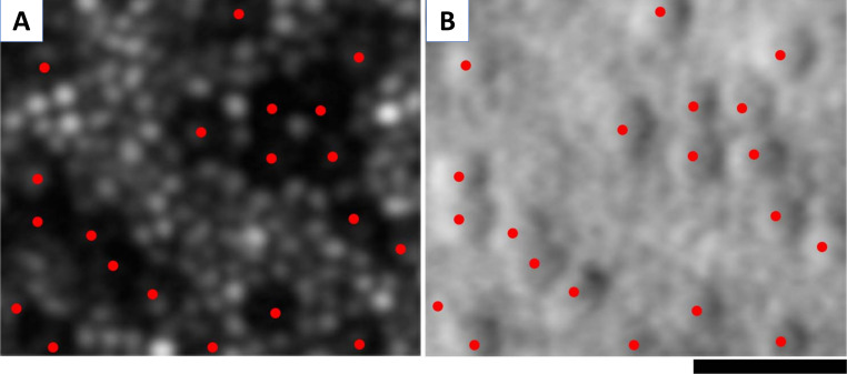
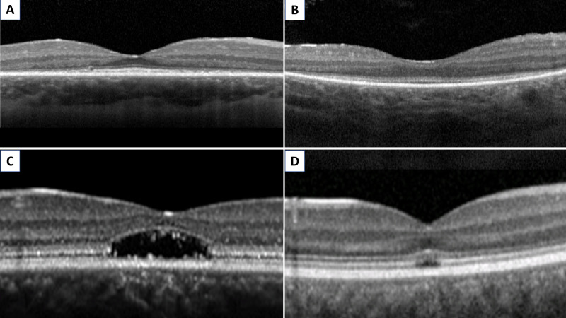
Ophthalmic genetics is a field that has been rapidly evolving over the last decade, mainly due to the flourishing of translational medicine for inherited retinal diseases (IRD). In this review, we will address the different methods by which retinal structure can be objectively and accurately assessed in IRD. We review standard-of-care imaging for these patients: colour fundus photography, fundus autofluorescence imaging and optical coherence tomography (OCT), as well as higher-resolution and/or newer technologies including OCT angiography, adaptive optics imaging, fundus imaging using a range of wavelengths, magnetic resonance imaging, laser speckle flowgraphy and retinal oximetry, illustrating their utility using paradigm genotypes with on-going therapeutic efforts/trials.

摘要

眼科遗传学是一个在过去十年中迅速发展的领域,主要得益于遗传性视网膜疾病(IRD)转化医学的兴起。在这篇综述中,我们将讨论评估 IRD 中视网膜结构的不同客观、准确的方法。我们回顾了这些患者的标准护理成像方法:彩色眼底照相、眼底自发荧光成像和光学相干断层扫描(OCT),以及包括 OCT 血管造影、自适应光学成像、使用多种波长的眼底成像、磁共振成像、激光散斑血流图和视网膜血氧测定等更高分辨率和/或更新的技术,并使用正在进行治疗努力/试验的范例基因型来说明它们的效用。

https://cdn.ncbi.nlm.nih.gov/pmc/blobs/9978/8639906/fe514dd0d745/bjophthalmol-2021-319228f03.jpg
https://cdn.ncbi.nlm.nih.gov/pmc/blobs/9978/8639906/e5f083ac7b13/bjophthalmol-2021-319228f01.jpg
https://cdn.ncbi.nlm.nih.gov/pmc/blobs/9978/8639906/1d0185b34a92/bjophthalmol-2021-319228f02.jpg
https://cdn.ncbi.nlm.nih.gov/pmc/blobs/9978/8639906/fe514dd0d745/bjophthalmol-2021-319228f03.jpg
https://cdn.ncbi.nlm.nih.gov/pmc/blobs/9978/8639906/e5f083ac7b13/bjophthalmol-2021-319228f01.jpg
https://cdn.ncbi.nlm.nih.gov/pmc/blobs/9978/8639906/1d0185b34a92/bjophthalmol-2021-319228f02.jpg
https://cdn.ncbi.nlm.nih.gov/pmc/blobs/9978/8639906/fe514dd0d745/bjophthalmol-2021-319228f03.jpg

相似文献

1
Structural evaluation in inherited retinal diseases.遗传性视网膜疾病的结构评估。
Br J Ophthalmol. 2021 Dec;105(12):1623-1631. doi: 10.1136/bjophthalmol-2021-319228. Epub 2021 May 12.
2
The retinal disease screening study: retrospective comparison of nonmydriatic fundus photography and three-dimensional optical coherence tomography for detection of retinal irregularities.视网膜疾病筛查研究:非散瞳眼底照相术与三维光相干断层扫描检测视网膜不规则性的回顾性比较。
Invest Ophthalmol Vis Sci. 2013 Aug 21;54(8):5694-700. doi: 10.1167/iovs.13-12043.
3
[New retinal imaging techniques].[新型视网膜成像技术]
Rev Prat. 2013 Jan;63(1):49-54.
4
In Vivo Imaging of Rodent Retina in Retinal Disease.在视网膜疾病中的啮齿动物视网膜的体内成像。
Methods Mol Biol. 2025;2848:151-167. doi: 10.1007/978-1-0716-4087-6_10.
5
Comparison of ultrahigh- and standard-resolution optical coherence tomography for imaging macular pathology.超高分辨率与标准分辨率光学相干断层扫描成像黄斑病变的比较。
Ophthalmology. 2005 Nov;112(11):1922.e1-15. doi: 10.1016/j.ophtha.2005.05.027. Epub 2005 Sep 23.
6
The retinal disease screening study: prospective comparison of nonmydriatic fundus photography and optical coherence tomography for detection of retinal irregularities.视网膜疾病筛查研究:非散瞳眼底照相术与光学相干断层扫描对视盘不规则性检测的前瞻性比较
Invest Ophthalmol Vis Sci. 2013 Feb 19;54(2):1460-8. doi: 10.1167/iovs.12-10727.
7
Acute Solar Retinopathy Imaged With Adaptive Optics, Optical Coherence Tomography Angiography, and En Face Optical Coherence Tomography.急性太阳视网膜病变的自适应光学成像、光相干断层扫描血管造影和共焦光学相干断层扫描。
JAMA Ophthalmol. 2018 Jan 1;136(1):82-85. doi: 10.1001/jamaophthalmol.2017.5517.
8
Current role of optical coherence tomography angiography: Expert panel discussion.光学相干断层扫描血管造影的当前作用:专家小组讨论。
Indian J Ophthalmol. 2018 Dec;66(12):1696-1699. doi: 10.4103/ijo.IJO_1048_18.
9
Multimodal imaging of Bietti's crystalline dystrophy.Bietti 结晶性营养不良的多模态成像。
Indian J Ophthalmol. 2018 Jul;66(7):1024-1026. doi: 10.4103/ijo.IJO_39_18.
10
Peripapillary Sparing in Autosomal Recessive Bestrophinopathy.常染色体隐性 Bestrophinopathy 中的视盘旁保留。
Ophthalmol Retina. 2020 May;4(5):523-529. doi: 10.1016/j.oret.2019.12.008. Epub 2020 Mar 5.

引用本文的文献

1
Retinograd-AI: An Open-Source Automated Fundus Autofluorescence Retinal Image Gradability Assessment for Inherited Retinal Diseases.视网膜图像分级人工智能:一种用于遗传性视网膜疾病的开源自动眼底自发荧光视网膜图像分级评估方法
Ophthalmol Sci. 2025 Jun 4;5(6):100845. doi: 10.1016/j.xops.2025.100845. eCollection 2025 Nov-Dec.
2
State of the Art on Inherited Retinal Dystrophies: Management and Molecular Genetics.遗传性视网膜营养不良的现状:管理与分子遗传学
J Clin Med. 2025 May 18;14(10):3526. doi: 10.3390/jcm14103526.
3
Discrepancies Between Autofluorescence Imaging Modalities in CNGB3-Associated Achromatopsia and Correlation With Ellipsoid Zone Continuity.

本文引用的文献

1
A demonstration of cone function plasticity after gene therapy in achromatopsia.基因治疗后天盲症中的锥体功能可塑性的演示。
Brain. 2022 Nov 21;145(11):3803-3815. doi: 10.1093/brain/awac226.
2
Retinal imaging in inherited retinal diseases.遗传性视网膜疾病中的视网膜成像。
Ann Eye Sci. 2020 Sep;5. doi: 10.21037/aes-20-81. Epub 2020 Sep 15.
3
Leber congenital amaurosis/early-onset severe retinal dystrophy: current management and clinical trials.Leber 先天性黑矇/早发性严重视网膜营养不良:当前的治疗方法和临床试验。
CNGB3相关性全色盲中自体荧光成像模式的差异及其与椭圆体区连续性的相关性
Transl Vis Sci Technol. 2025 May 1;14(5):24. doi: 10.1167/tvst.14.5.24.
4
Retinal Thickness Analysis Using Optical Coherence Tomography: Diagnostic and Monitoring Applications in Retinal Diseases.使用光学相干断层扫描的视网膜厚度分析:在视网膜疾病中的诊断与监测应用
Diagnostics (Basel). 2025 Mar 25;15(7):833. doi: 10.3390/diagnostics15070833.
5
Quantification of Fundus Autofluorescence Features in a Molecularly Characterized Cohort of >3500 Patients with Inherited Retinal Disease from the United Kingdom.对来自英国的3500多名遗传性视网膜疾病患者的分子特征队列中的眼底自发荧光特征进行量化。
Ophthalmol Sci. 2024 Nov 12;5(2):100652. doi: 10.1016/j.xops.2024.100652. eCollection 2025 Mar-Apr.
6
Utility of multimodal imaging in the clinical diagnosis of inherited retinal degenerations.多模态成像在遗传性视网膜变性临床诊断中的应用
Taiwan J Ophthalmol. 2024 Dec 3;14(4):486-496. doi: 10.4103/tjo.TJO-D-24-00066. eCollection 2024 Oct-Dec.
7
Update on Clinical Trial Endpoints in Gene Therapy Trials for Inherited Retinal Diseases.遗传性视网膜疾病基因治疗试验的临床试验终点更新
J Clin Med. 2024 Sep 18;13(18):5512. doi: 10.3390/jcm13185512.
8
IQCB1 (NPHP5)-Retinopathy: Clinical and Genetic Characterization and Natural History.IQCB1(NPHP5)相关性视网膜病变:临床与遗传学特征及自然病史
Am J Ophthalmol. 2024 Aug;264:205-215. doi: 10.1016/j.ajo.2024.03.009. Epub 2024 Mar 23.
9
Adaptive Optics Retinal Imaging in RDH12-Associated Early Onset Severe Retinal Dystrophy.RDH12相关早发性严重视网膜营养不良的自适应光学视网膜成像
Invest Ophthalmol Vis Sci. 2024 Mar 5;65(3):9. doi: 10.1167/iovs.65.3.9.
10
Spectrum of Genetic Variants in the Most Common Genes Causing Inherited Retinal Disease in a Large Molecularly Characterized United Kingdom Cohort.在一个经过大规模分子特征分析的英国队列中,最常见的遗传性视网膜疾病相关基因的遗传变异谱。
Ophthalmol Retina. 2024 Jul;8(7):699-709. doi: 10.1016/j.oret.2024.01.012. Epub 2024 Jan 12.
Br J Ophthalmol. 2022 Apr;106(4):445-451. doi: 10.1136/bjophthalmol-2020-318483. Epub 2021 Mar 12.
4
Quantitative Color Fundus Autofluorescence in Patients with Diabetes Mellitus.糖尿病患者的定量彩色眼底自发荧光
J Clin Med. 2020 Dec 25;10(1):48. doi: 10.3390/jcm10010048.
5
Deep Learning-Based Classification of Inherited Retinal Diseases Using Fundus Autofluorescence.基于深度学习的眼底自发荧光对遗传性视网膜疾病的分类
J Clin Med. 2020 Oct 14;9(10):3303. doi: 10.3390/jcm9103303.
6
Functional optical coherence tomography enables optoretinography of photoreceptor dysfunction due to retinal degeneration.功能性光学相干断层扫描能够对因视网膜变性导致的光感受器功能障碍进行视网膜电图检查。
Biomed Opt Express. 2020 Aug 27;11(9):5306-5320. doi: 10.1364/BOE.399334. eCollection 2020 Sep 1.
7
Intraobserver Repeatability and Interobserver Reproducibility of Foveal Cone Density Measurements in and -Associated Achromatopsia.中央凹视锥细胞密度测量在与疾病相关的全色盲中的观察者内重复性和观察者间再现性
Transl Vis Sci Technol. 2020 Jun 26;9(7):37. doi: 10.1167/tvst.9.7.37. eCollection 2020 Jun.
8
Deep Learning-Based Detection of Pigment Signs for Analysis and Diagnosis of Retinitis Pigmentosa.基于深度学习的色素征检测在分析和诊断色素性视网膜炎中的应用。
Sensors (Basel). 2020 Jun 18;20(12):3454. doi: 10.3390/s20123454.
9
The qualitative assessment of optical coherence tomography and the central retinal sensitivity in patients with retinitis pigmentosa.视网膜色素变性患者的光学相干断层扫描和中心视网膜敏感度的定性评估。
PLoS One. 2020 May 11;15(5):e0232700. doi: 10.1371/journal.pone.0232700. eCollection 2020.
10
Retinal Structure in RPE65-Associated Retinal Dystrophy.RPE65 相关视网膜营养不良的视网膜结构。
Invest Ophthalmol Vis Sci. 2020 Apr 9;61(4):47. doi: 10.1167/iovs.61.4.47.