Komada Tomohiro, Suzuki Kojiro, Ishiguchi Hiroaki, Kawai Hisashi, Okumura Takahiro, Hirashiki Akihiro, Naganawa Shinji
Department of Radiology, Nagoya University Graduate School of Medicine, Nagoya, Japan.
Department of Advanced Medicine in Cardiopulmonary Disease, Nagoya University Graduate School of Medicine, Nagoya, Japan.
Nagoya J Med Sci. 2016 Dec;78(4):437-446. doi: 10.18999/nagjms.78.4.437.
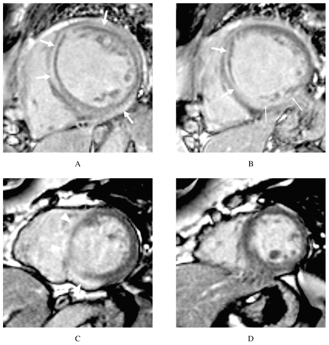
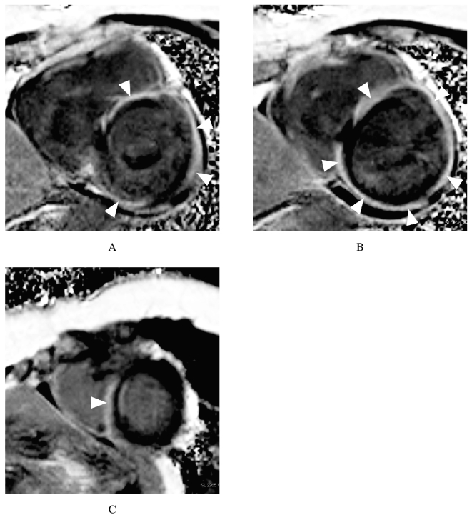
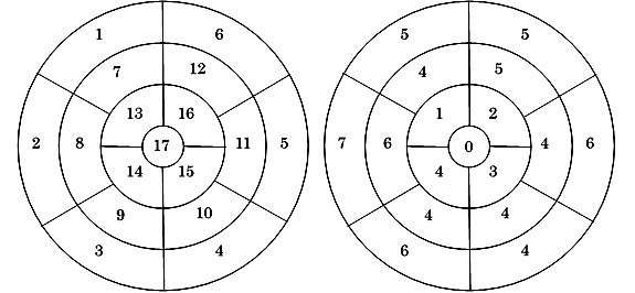
Cardiac sarcoidosis (CS) can cause sudden death, which is the leading cause of mortality in patients with sarcoidosis in Japan. However, it is difficult to diagnose CS because of the lack of a sensitive diagnostic method for the condition. Late gadolinium-enhanced cardiac magnetic resonance (MR) imaging demonstrates improved sensitivity for diagnosing CS. Therefore, it is important to know the late gadolinium-enhancement (LGE) characteristics of CS on cardiac MR images in order to diagnose CS accurately. In this study, we investigated the most common sites of LGE on cardiac MR images in CS. Late gadolinium-enhanced MR images of 9 consecutive patients with CS (obtained between August 2009 and July 2015) were reviewed by two radiologists. The distribution of LGE was evaluated using the American Heart Association 17-segment model of the left ventricle. The LGE in each segment was also classified into 4 patterns according to the myocardial layer in which it occurred (the subepicardial, subendocardial, intramural, and transmural layer patterns). All 9 patients exhibited LGE in their left ventricle, and 70 of 153 (46%) myocardial segments were enhanced. All of the patients displayed LGE in the basal septal wall. The patients' LGE layer patterns were as follows: subepicardial: 40% (28/70), intramural: 30% (21/70), subendocardial: 16% (11/70), and transmural: 14% (10/70). The basal septum wall and subepicardial layer often exhibit LGE on cardiac MR images in CS patients. LGE can be observed in other segments and layers in some cases.
心脏结节病(CS)可导致猝死,这是日本结节病患者死亡的主要原因。然而,由于缺乏针对该病的敏感诊断方法,CS很难被诊断出来。延迟钆增强心脏磁共振(MR)成像对CS诊断的敏感性有所提高。因此,了解CS在心脏MR图像上的延迟钆增强(LGE)特征对于准确诊断CS很重要。在本研究中,我们调查了CS患者心脏MR图像上LGE最常见的部位。两名放射科医生对9例连续的CS患者(于2009年8月至2015年7月期间获得)的延迟钆增强MR图像进行了回顾。使用美国心脏协会的左心室17节段模型评估LGE的分布。每个节段的LGE也根据其发生的心肌层分为4种模式(心外膜下、心内膜下、壁内和透壁层模式)。所有9例患者左心室均出现LGE,153个心肌节段中有70个(46%)出现强化。所有患者的基底间隔壁均出现LGE。患者的LGE层模式如下:心外膜下:40%(28/70),壁内:30%(21/70),心内膜下:16%(11/70),透壁:14%(10/70)。CS患者心脏MR图像上,基底间隔壁和心外膜下层常出现LGE。在某些情况下,其他节段和层也可观察到LGE。