• 文献检索
  • 文档翻译
  • 深度研究
  • 学术资讯
  • Suppr Zotero 插件Zotero 插件
  • 邀请有礼
  • 套餐&价格
  • 历史记录
应用&插件
Suppr Zotero 插件Zotero 插件浏览器插件Mac 客户端Windows 客户端微信小程序
定价
高级版会员购买积分包购买API积分包
服务
文献检索文档翻译深度研究API 文档MCP 服务
关于我们
关于 Suppr公司介绍联系我们用户协议隐私条款
关注我们

Suppr 超能文献

核心技术专利:CN118964589B侵权必究
粤ICP备2023148730 号-1Suppr @ 2026

文献检索

告别复杂PubMed语法,用中文像聊天一样搜索,搜遍4000万医学文献。AI智能推荐,让科研检索更轻松。

立即免费搜索

文件翻译

保留排版,准确专业,支持PDF/Word/PPT等文件格式,支持 12+语言互译。

免费翻译文档

深度研究

AI帮你快速写综述,25分钟生成高质量综述,智能提取关键信息,辅助科研写作。

立即免费体验

用于在小角X射线散射实验中自动进行辐射损伤定量分析的工具的开发。

Development of tools to automate quantitative analysis of radiation damage in SAXS experiments.

作者信息

Brooks-Bartlett Jonathan C, Batters Rebecca A, Bury Charles S, Lowe Edward D, Ginn Helen Mary, Round Adam, Garman Elspeth F

机构信息

Department of Biochemistry, University of Oxford, Oxford OX1 3QU, UK.

Division of Structural Biology, Wellcome Trust Centre for Human Genetics, Roosevelt Drive, Oxford OX3 7BN, UK.

出版信息

J Synchrotron Radiat. 2017 Jan 1;24(Pt 1):63-72. doi: 10.1107/S1600577516015083.

DOI:10.1107/S1600577516015083
PMID:28009547
原文链接:https://pmc.ncbi.nlm.nih.gov/articles/PMC5182020/
Abstract

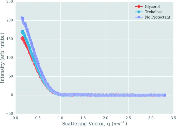
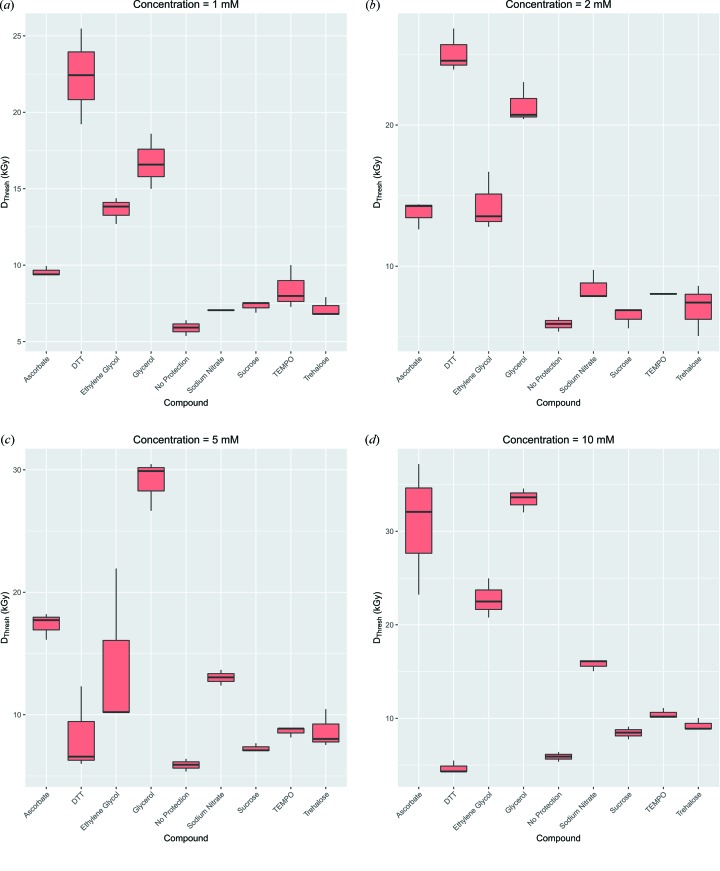
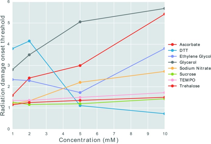
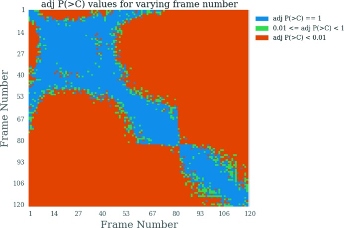
Biological small-angle X-ray scattering (SAXS) is an increasingly popular technique used to obtain nanoscale structural information on macromolecules in solution. However, radiation damage to the samples limits the amount of useful data that can be collected from a single sample. In contrast to the extensive analytical resources available for macromolecular crystallography (MX), there are relatively few tools to quantitate radiation damage for SAXS, some of which require a significant level of manual characterization, with the potential of leading to conflicting results from different studies. Here, computational tools have been developed to automate and standardize radiation damage analysis for SAXS data. RADDOSE-3D, a dose calculation software utility originally written for MX experiments, has been extended to account for the cylindrical geometry of the capillary tube, the liquid composition of the sample and the attenuation of the beam by the capillary material to allow doses to be calculated for many SAXS experiments. Furthermore, a library has been written to visualize and explore the pairwise similarity of frames. The calculated dose for the frame at which three subsequent frames are determined to be dissimilar is defined as the radiation damage onset threshold (RDOT). Analysis of RDOTs has been used to compare the efficacy of radioprotectant compounds to extend the useful lifetime of SAXS samples. Comparison of the RDOTs shows that, for radioprotectant compounds at 5 and 10 mM concentration, glycerol is the most effective compound. However, at 1 and 2 mM concentrations, dithiothreitol (DTT) appears to be most effective. Our newly developed visualization library contains methods that highlight the unusual radiation damage results given by SAXS data collected using higher concentrations of DTT: these observations should pave the way to the development of more sophisticated frame merging strategies.

摘要

生物小角X射线散射(SAXS)是一种越来越受欢迎的技术,用于获取溶液中大分子的纳米级结构信息。然而,样品的辐射损伤限制了从单个样品中可收集的有用数据量。与大分子晶体学(MX)可用的大量分析资源相比,用于定量SAXS辐射损伤的工具相对较少,其中一些需要大量的手动表征,可能导致不同研究结果相互矛盾。在这里,已经开发了计算工具来自动化和标准化SAXS数据的辐射损伤分析。RADDOSE-3D是一个最初为MX实验编写的剂量计算软件实用程序,已扩展到考虑毛细管的圆柱形几何形状、样品的液体成分以及毛细管材料对光束的衰减,以便为许多SAXS实验计算剂量。此外,还编写了一个库来可视化和探索帧的成对相似性。将确定三个后续帧不同的帧的计算剂量定义为辐射损伤起始阈值(RDOT)。对RDOT的分析已用于比较辐射防护化合物延长SAXS样品使用寿命的功效。RDOT的比较表明,对于浓度为5和10 mM的辐射防护化合物,甘油是最有效的化合物。然而,在浓度为1和2 mM时,二硫苏糖醇(DTT)似乎最有效。我们新开发的可视化库包含一些方法,这些方法突出了使用较高浓度DTT收集的SAXS数据给出的异常辐射损伤结果:这些观察结果应该为开发更复杂的帧合并策略铺平道路

https://cdn.ncbi.nlm.nih.gov/pmc/blobs/25c1/5182020/6ae2da96e0bd/s-24-00063-fig9.jpg
https://cdn.ncbi.nlm.nih.gov/pmc/blobs/25c1/5182020/d4399b15b447/s-24-00063-fig1.jpg
https://cdn.ncbi.nlm.nih.gov/pmc/blobs/25c1/5182020/76ac6a836d5e/s-24-00063-fig2.jpg
https://cdn.ncbi.nlm.nih.gov/pmc/blobs/25c1/5182020/6e611374904a/s-24-00063-fig3.jpg
https://cdn.ncbi.nlm.nih.gov/pmc/blobs/25c1/5182020/67762863f335/s-24-00063-fig4.jpg
https://cdn.ncbi.nlm.nih.gov/pmc/blobs/25c1/5182020/6cf2f37f8189/s-24-00063-fig5.jpg
https://cdn.ncbi.nlm.nih.gov/pmc/blobs/25c1/5182020/989d5524168c/s-24-00063-fig6.jpg
https://cdn.ncbi.nlm.nih.gov/pmc/blobs/25c1/5182020/16598c014647/s-24-00063-fig7.jpg
https://cdn.ncbi.nlm.nih.gov/pmc/blobs/25c1/5182020/521f03700f6b/s-24-00063-fig8.jpg
https://cdn.ncbi.nlm.nih.gov/pmc/blobs/25c1/5182020/6ae2da96e0bd/s-24-00063-fig9.jpg

相似文献

1
Development of tools to automate quantitative analysis of radiation damage in SAXS experiments.用于在小角X射线散射实验中自动进行辐射损伤定量分析的工具的开发。
J Synchrotron Radiat. 2017 Jan 1;24(Pt 1):63-72. doi: 10.1107/S1600577516015083.
2
Smaller capillaries improve the small-angle X-ray scattering signal and sample consumption for biomacromolecular solutions.较小的毛细管可改善生物大分子溶液的小角X射线散射信号和样品消耗。
J Synchrotron Radiat. 2018 Jul 1;25(Pt 4):1113-1122. doi: 10.1107/S1600577518007907. Epub 2018 Jun 26.
3
Structural Modeling Using Solution Small-Angle X-ray Scattering (SAXS).利用溶液小角 X 射线散射(SAXS)进行结构建模。
J Mol Biol. 2020 Apr 17;432(9):3078-3092. doi: 10.1016/j.jmb.2020.01.030. Epub 2020 Feb 6.
4
X-ray radiation damage to biological macromolecules: further insights.X射线对生物大分子的辐射损伤:进一步的见解
J Synchrotron Radiat. 2017 Jan 1;24(Pt 1):1-6. doi: 10.1107/S160057751602018X.
5
Doses for experiments with microbeams and microcrystals: Monte Carlo simulations in RADDOSE-3D.用于微束和微晶体实验的剂量:RADDOSE-3D 中的蒙特卡罗模拟。
Protein Sci. 2021 Jan;30(1):8-19. doi: 10.1002/pro.3922. Epub 2020 Aug 18.
6
Limiting radiation damage for high-brilliance biological solution scattering: practical experience at the EMBL P12 beamline PETRAIII.限制高亮度生物溶液散射的辐射损伤:欧洲分子生物学实验室P12光束线PETRAIII的实践经验。
J Synchrotron Radiat. 2015 Mar;22(2):273-9. doi: 10.1107/S1600577515000375. Epub 2015 Feb 4.
7
Evolving SAXS versatility: solution X-ray scattering for macromolecular architecture, functional landscapes, and integrative structural biology.不断发展的小角 X 射线散射技术的多功能性:用于大分子结构、功能景观和综合结构生物学的溶液 X 射线散射技术。
Curr Opin Struct Biol. 2019 Oct;58:197-213. doi: 10.1016/j.sbi.2019.04.004. Epub 2019 Jun 13.
8
Uridine as a new scavenger for synchrotron-based structural biology techniques.尿苷作为基于同步加速器的结构生物学技术的新型清除剂。
J Synchrotron Radiat. 2017 Jan 1;24(Pt 1):53-62. doi: 10.1107/S1600577516018452.
9
Advances in X-ray scattering: from solution SAXS to achievements with coherent beams.X 射线散射的进展:从溶液 SAXS 到相干光束的成就。
Curr Opin Struct Biol. 2012 Oct;22(5):670-8. doi: 10.1016/j.sbi.2012.07.014. Epub 2012 Sep 3.
10
Visualizing and accessing correlated SAXS data sets with Similarity Maps and Simple Scattering web resources.利用相似性图谱和简单散射网络资源对相关 SAXS 数据集进行可视化和访问。
Methods Enzymol. 2023;678:411-440. doi: 10.1016/bs.mie.2022.09.024. Epub 2022 Nov 3.

引用本文的文献

1
AF4-to-SAXS: expanded characterization of nanoparticles and proteins at the P12 BioSAXS beamline.从不对称流场分离到小角X射线散射:在P12生物小角X射线散射光束线对纳米颗粒和蛋白质进行扩展表征
J Synchrotron Radiat. 2025 Jul 1;32(Pt 4):971-985. doi: 10.1107/S1600577525003959. Epub 2025 Jun 12.
2
SAXS measurements of azobenzene lipid vesicles reveal buffer-dependent photoswitching and quantitative isomerisation by X-rays.偶氮苯脂质囊泡的小角X射线散射测量揭示了缓冲液依赖性光开关效应以及X射线诱导的定量异构化。
Nanophotonics. 2022 Apr 15;11(10):2361-2368. doi: 10.1515/nanoph-2022-0053. eCollection 2022 May.
3
Doses for X-ray and electron diffraction: New features in RADDOSE-3D including intensity decay models.

本文引用的文献

1
Quantifying radiation damage in biomolecular small-angle X-ray scattering.量化生物分子小角X射线散射中的辐射损伤。
J Appl Crystallogr. 2016 May 4;49(Pt 3):880-890. doi: 10.1107/S1600576716005136. eCollection 2016 Jun 1.
2
A Brief Survey of State-of-the-Art BioSAXS.最新生物小角X射线散射技术概述
Protein Pept Lett. 2016;23(3):217-31. doi: 10.2174/0929866523666160106153655.
3
A microfabricated fixed path length silicon sample holder improves background subtraction for cryoSAXS.一种微制造的固定路径长度硅样品架改善了低温小角X射线散射的背景扣除。
X 射线和电子衍射剂量:RADDOSE-3D 中的新功能,包括强度衰减模型。
Protein Sci. 2024 Jul;33(7):e5005. doi: 10.1002/pro.5005.
4
Adaptable SEC-SAXS data collection for higher quality structure analysis in solution.适应力强的 SEC-SAXS 数据采集可提高溶液中结构分析的质量。
Protein Sci. 2024 Apr;33(4):e4946. doi: 10.1002/pro.4946.
5
Lattice response to the radiation damage of molecular crystals: radiation-induced versus thermal expansivity.晶格对分子晶体辐射损伤的响应:辐射诱导与热膨胀率
Acta Crystallogr B Struct Sci Cryst Eng Mater. 2024 Feb 1;80(Pt 1):13-18. doi: 10.1107/S2052520623010636. Epub 2024 Jan 4.
6
Variability in X-ray induced effects in [Rh(COD)Cl] with changing experimental parameters.随着实验参数的变化,[Rh(COD)Cl]中 X 射线诱导效应的变化。
Phys Chem Chem Phys. 2022 Nov 30;24(46):28444-28456. doi: 10.1039/d2cp03928a.
7
A setup for millisecond time-resolved X-ray solution scattering experiments at the CoSAXS beamline at the MAX IV Laboratory.在 MAX IV 实验室的 CoSAXS 光束线上进行毫秒时间分辨 X 射线溶液散射实验的设置。
J Synchrotron Radiat. 2022 Mar 1;29(Pt 2):555-562. doi: 10.1107/S1600577522000996. Epub 2022 Feb 16.
8
Negative X-ray expansion in cadmium cyanide.氰化镉中的负X射线膨胀。
Mater Horiz. 2021 May 1;8(5):1446-1453. doi: 10.1039/d0mh01989e. Epub 2021 Feb 23.
9
A SAXS-based approach to rationally evaluate radical scavengers - toward eliminating radiation damage in solution and crystallographic studies.基于小角 X 射线散射的方法来合理评估自由基清除剂 - 旨在消除溶液和晶体学研究中的辐射损伤。
J Synchrotron Radiat. 2021 Sep 1;28(Pt 5):1309-1320. doi: 10.1107/S1600577521004045. Epub 2021 Aug 9.
10
Radiation damage to biological samples: still a pertinent issue.生物样本的辐射损伤:仍是一个相关问题。
J Synchrotron Radiat. 2021 Sep 1;28(Pt 5):1278-1283. doi: 10.1107/S1600577521008845.
J Appl Crystallogr. 2015 Jan 30;48(Pt 1):227-237. doi: 10.1107/S1600576714027782. eCollection 2015 Feb 1.
4
A micro-patterned silicon chip as sample holder for macromolecular crystallography experiments with minimal background scattering.一种微图案化硅芯片,用作大分子晶体学实验的样品架,背景散射极小。
Sci Rep. 2015 May 29;5:10451. doi: 10.1038/srep10451.
5
Correlation Map, a goodness-of-fit test for one-dimensional X-ray scattering spectra.关联图,一种用于一维 X 射线散射光谱的拟合优度检验。
Nat Methods. 2015 May;12(5):419-22. doi: 10.1038/nmeth.3358. Epub 2015 Apr 6.
6
Versatile sample environments and automation for biological solution X-ray scattering experiments at the P12 beamline (PETRA III, DESY).用于P12光束线(德国电子同步加速器研究所的PETRA III)生物溶液X射线散射实验的多功能样品环境和自动化技术。
J Appl Crystallogr. 2015 Mar 12;48(Pt 2):431-443. doi: 10.1107/S160057671500254X. eCollection 2015 Apr 1.
7
Exploring oxidative modifications of tyrosine: an update on mechanisms of formation, advances in analysis and biological consequences.探索酪氨酸的氧化修饰:形成机制的最新进展、分析方法的进步及生物学后果
Free Radic Res. 2015 Apr;49(4):347-73. doi: 10.3109/10715762.2015.1007968. Epub 2015 Mar 27.
8
Limiting radiation damage for high-brilliance biological solution scattering: practical experience at the EMBL P12 beamline PETRAIII.限制高亮度生物溶液散射的辐射损伤:欧洲分子生物学实验室P12光束线PETRAIII的实践经验。
J Synchrotron Radiat. 2015 Mar;22(2):273-9. doi: 10.1107/S1600577515000375. Epub 2015 Feb 4.
9
Diamond Light Source: status and perspectives.钻石光源:现状与展望。
Philos Trans A Math Phys Eng Sci. 2015 Mar 6;373(2036). doi: 10.1098/rsta.2013.0161.
10
BioSAXS Sample Changer: a robotic sample changer for rapid and reliable high-throughput X-ray solution scattering experiments.小角X射线散射样品更换器:一种用于快速且可靠的高通量X射线溶液散射实验的自动样品更换器。
Acta Crystallogr D Biol Crystallogr. 2015 Jan 1;71(Pt 1):67-75. doi: 10.1107/S1399004714026959.