Patil Savita S, Kudrimoti Jyoti K, Agarwal Rachana D, Jadhav Meenal V, Chuge Ashish
Department of Pathology and Neurosurgery, B J Government Medical College, Pune, Maharashtra, India.
J Cytol. 2016 Oct-Dec;33(4):205-209. doi: 10.4103/0970-9371.190442.
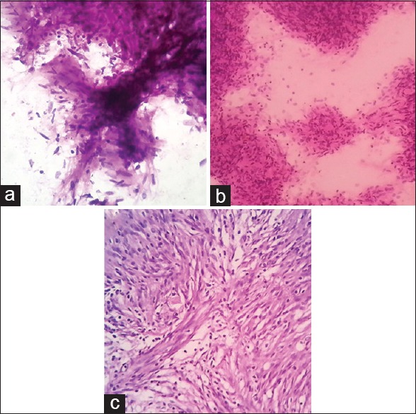
Central nervous system (CNS) squash cytology (CSC) has established itself as a technically simple, rapid, inexpensive, fairly accurate, and dependable intraoperative diagnostic tool. It helps neurosurgeons immensely when management is dependent on it.
This study aimed at finding out the utility of CSC as an intraoperative diagnostic tool from a neurosurgeon's perspective.
Fifty prospectively registered patients with clinical diagnosis of CNS tumors were enrolled in the study. All the patients were subjected to magnetic resonance imaging (MRI). Intraoperative CSC was performed and smears were stained with Leishman and rapid Hematoxylin and Eosin (H and E) stain. The diagnosis of CSC was compared with MRI diagnosis and histopathological diagnosis. The CNS tumors were categorized based on clinical and therapeutic implications. Diagnostic accuracy, sensitivity, specificity, and positive and negative predictive value of MRI and CSC were calculated by using appropriate formulae.
The age range of the CNS tumors included in the study was 2 to 68 years. There was a slight female preponderance. Sensitivity, specificity, positive predictive value, and negative predictive value of preoperative MRI were 90.47%, 82.76%, 79.17%, and 92.31% respectively. These values of utility parameters for CSC were 100% for each of the clinical and therapeutic implications. It helped neurosurgeons in optimizing surgical procedure in 12 cases of meningioma. It influenced surgical management in 1 case of infratentorial pilocytic astrocytoma, and helped in the diagnosis and management of 9 unexpected tumors missed on MRI.
中枢神经系统(CNS)挤压细胞学检查(CSC)已成为一种技术简单、快速、廉价、相当准确且可靠的术中诊断工具。当治疗依赖于它时,它对神经外科医生有极大帮助。
本研究旨在从神经外科医生的角度探讨CSC作为术中诊断工具的实用性。
本研究纳入了50例经临床诊断为中枢神经系统肿瘤的前瞻性登记患者。所有患者均接受了磁共振成像(MRI)检查。术中进行CSC检查,并将涂片用利什曼染色以及快速苏木精和伊红(H&E)染色。将CSC诊断结果与MRI诊断结果和组织病理学诊断结果进行比较。根据临床和治疗意义对中枢神经系统肿瘤进行分类。使用适当公式计算MRI和CSC的诊断准确性、敏感性、特异性以及阳性和阴性预测值。
本研究纳入的中枢神经系统肿瘤患者年龄范围为2至68岁。女性略占优势。术前MRI的敏感性、特异性、阳性预测值和阴性预测值分别为90.47%、82.76%、79.17%和92.31%。CSC的这些实用参数值在每种临床和治疗意义方面均为100%。它帮助神经外科医生在12例脑膜瘤手术中优化了手术操作。它影响了1例幕下毛细胞型星形细胞瘤的手术治疗,并帮助诊断和处理了9例MRI漏诊的意外肿瘤。