Altieri Barbara, Sbiera Silviu, Della Casa Silvia, Weigand Isabel, Wild Vanessa, Steinhauer Sonja, Fadda Guido, Kocot Arkadius, Bekteshi Michaela, Mambretti Egle M, Rosenwald Andreas, Pontecorvi Alfredo, Fassnacht Martin, Ronchi Cristina L
Department of Internal Medicine I, Division of Endocrinology and Diabetes, University Hospital of Wuerzburg, Germany.
Division of Endocrinology and Metabolic Diseases, Catholic University of the Sacred Heart, Rome, Italy.
Oncotarget. 2017 Feb 7;8(6):9323-9338. doi: 10.18632/oncotarget.14067.
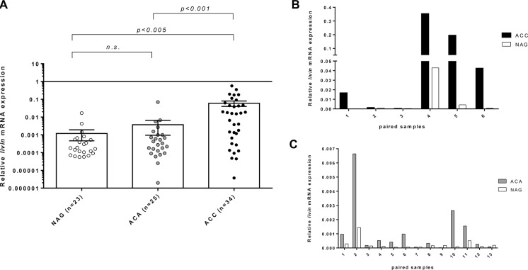
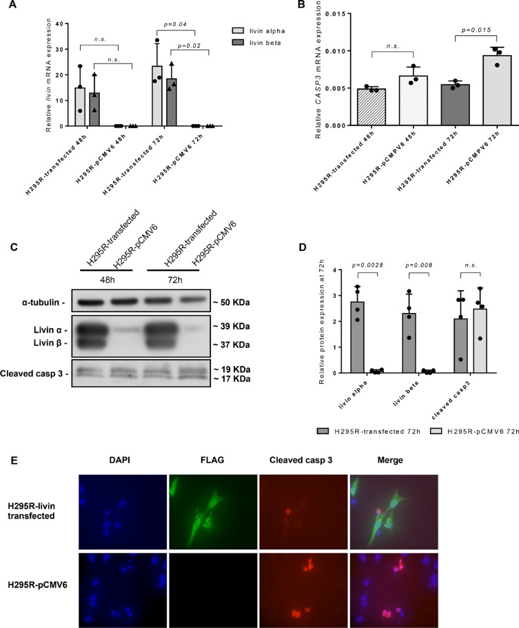
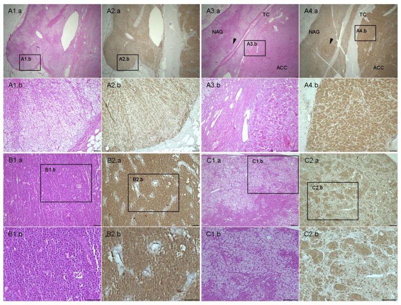
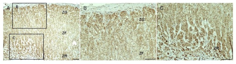
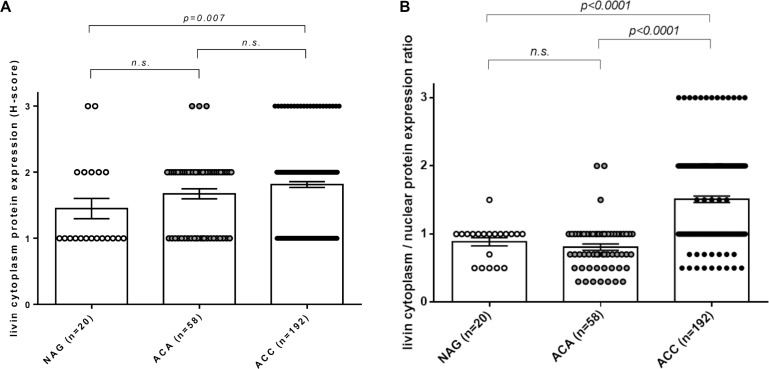
Livin/BIRC7 is a member of the inhibitors of apoptosis proteins family, which are involved in tumor development through the inhibition of caspases. Aim was to investigate the expression of livin and other members of its pathway in adrenocortical tumors and in the adrenocortical carcinoma (ACC) cell line NCI-H295R.The mRNA expression of livin, its isoforms α and β, XIAP, CASP3 and DIABLO was evaluated by qRT-PCR in 82 fresh-frozen adrenal tissues (34 ACC, 25 adenomas = ACA, 23 normal adrenal glands = NAG). Livin protein expression was assessed by immunohistochemistry in 270 paraffin-embedded tissues (192 ACC, 58 ACA, 20 NAG). Livin, CASP3 and cleaved caspase-3 were evaluated in NCI-H295R after induction of livin overexpression.Relative livin mRNA expression was significantly higher in ACC than in ACA and NAG (0.060 ± 0.116 vs 0.004 ± 0.014 and 0.002 ± 0.009, respectively, p < 0.01), being consistently higher in tumors than in adjacent NAG and isoform β more expressed than α. No significant differences in CASP3, XIAP and DIABLO levels were found among these groups. In immunohistochemistry, livin was localized in both cytoplasm and nuclei. The ratio between cytoplasmic and nuclear staining was significantly higher in ACC (1.51 ± 0.66) than in ACA (0.80 ± 0.35) and NAG (0.88 ± 0.27; p < 0.0001). No significant correlations were observed between livin expression and histopathological parameters or clinical outcome. In NCI-H295R cells, the livin overexpression slightly reduced the activation of CASP3, but did not correlate with cell viability.In conclusion, livin is specifically over-expressed in ACC, suggesting that it might be involved in adrenocortical tumorigenesis and represent a new molecular marker of malignancy.
生存素(Livin)/BIRC7是凋亡抑制蛋白家族的成员之一,该家族通过抑制半胱天冬酶参与肿瘤发展。目的是研究生存素及其通路其他成员在肾上腺皮质肿瘤和肾上腺皮质癌(ACC)细胞系NCI-H295R中的表达情况。通过qRT-PCR评估了82个新鲜冷冻肾上腺组织(34例ACC、25例腺瘤=ACA、23例正常肾上腺=NAG)中生存素及其α和β亚型、XIAP、CASP3和DIABLO的mRNA表达。通过免疫组织化学评估了270个石蜡包埋组织(192例ACC、58例ACA、20例NAG)中生存素蛋白的表达。在诱导生存素过表达后,对NCI-H295R细胞中的生存素、CASP3和裂解的半胱天冬酶-3进行了评估。ACC中生存素相对mRNA表达显著高于ACA和NAG(分别为0.060±0.116 vs 0.004±0.014和0.002±0.009,p<0.01),肿瘤中的表达始终高于相邻的NAG,且β亚型的表达高于α亚型。这些组之间在CASP3、XIAP和DIABLO水平上未发现显著差异。在免疫组织化学中,生存素定位于细胞质和细胞核中。ACC中细胞质与细胞核染色的比例(1.51±0.66)显著高于ACA(0.80±0.35)和NAG(0.88±0.27;p<0.0001)。生存素表达与组织病理学参数或临床结果之间未观察到显著相关性。在NCI-H295R细胞中,生存素过表达略微降低了CASP3的激活,但与细胞活力无关。总之,生存素在ACC中特异性过表达,提示其可能参与肾上腺皮质肿瘤发生,并且是一种新的恶性分子标志物。